 Movie
Movie Controller

[English] 日本語
 Yorodumi
- PDB-5tre: Zinc and the Iron Donor Frataxin Regulate Oligomerization of the ... -
+ Open data

- Basic information
| Entry | Database: PDB / ID: 5tre | ||||||
|---|---|---|---|---|---|---|---|
| Title | Zinc and the Iron Donor Frataxin Regulate Oligomerization of the Scaffold Protein to Form New Fe-S Cluster Assembly Centers | ||||||
 Components Components |
| ||||||
 Keywords Keywords | OXIDOREDUCTASE / Friedreich Ataxia / frataxin / iron-sulfur protein / mitochondria / protein complex | ||||||
| Function / homology |  Function and homology information Function and homology informationMitochondrial iron-sulfur cluster biogenesis / Mitochondrial protein import / Maturation of TCA enzymes and regulation of TCA cycle / Complex III assembly / mitochondrial electron transport, succinate to ubiquinone / tRNA wobble uridine modification / iron chaperone activity / iron-sulfur cluster assembly complex / heme biosynthetic process / iron-sulfur cluster assembly ...Mitochondrial iron-sulfur cluster biogenesis / Mitochondrial protein import / Maturation of TCA enzymes and regulation of TCA cycle / Complex III assembly / mitochondrial electron transport, succinate to ubiquinone / tRNA wobble uridine modification / iron chaperone activity / iron-sulfur cluster assembly complex / heme biosynthetic process / iron-sulfur cluster assembly / response to iron(II) ion / ferroxidase / ATPase activator activity / ferroxidase activity / ferric iron binding / glutathione metabolic process / iron ion transport / ferrous iron binding / mitochondrial intermembrane space / 2 iron, 2 sulfur cluster binding / response to oxidative stress / intracellular iron ion homeostasis / mitochondrial inner membrane / iron ion binding / mitochondrial matrix / mitochondrion / zinc ion binding / identical protein binding / cytoplasm Similarity search - Function | ||||||
| Biological species |  | ||||||
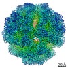
| Method | ELECTRON MICROSCOPY / single particle reconstruction / negative staining / Resolution: 15.6 Å | ||||||
 Authors Authors | Ranatunga, W. / Gakh, O. / Galeano, B.K. / Smith IV, D.Y. / Thompson, J.R. / Isaya, G. | ||||||
| Funding support |  United States, 1items United States, 1items
| ||||||
 Citation Citation | Journal: Metallomics / Year: 2017 Title: Zinc and the iron donor frataxin regulate oligomerization of the scaffold protein to form new Fe-S cluster assembly centers. Authors: B K Galeano / W Ranatunga / O Gakh / D Y Smith / J R Thompson / G Isaya / Abstract: Early studies of the bacterial Fe-S cluster assembly system provided structural details for how the scaffold protein and the cysteine desulfurase interact. This work and additional work on the yeast ...Early studies of the bacterial Fe-S cluster assembly system provided structural details for how the scaffold protein and the cysteine desulfurase interact. This work and additional work on the yeast and human systems elucidated a conserved mechanism for sulfur donation but did not provide any conclusive insights into the mechanism for iron delivery from the iron donor, frataxin, to the scaffold. We previously showed that oligomerization is a mechanism by which yeast frataxin (Yfh1) can promote assembly of the core machinery for Fe-S cluster synthesis both in vitro and in cells, in such a manner that the scaffold protein, Isu1, can bind to Yfh1 independent of the presence of the cysteine desulfurase, Nfs1. Here, in the absence of Yfh1, Isu1 was found to exist in two forms, one mostly monomeric with limited tendency to dimerize, and one with a strong propensity to oligomerize. Whereas the monomeric form is stabilized by zinc, the loss of zinc promotes formation of dimer and higher order oligomers. However, upon binding to oligomeric Yfh1, both forms take on a similar symmetrical trimeric configuration that places the Fe-S cluster coordinating residues of Isu1 in close proximity of iron-binding residues of Yfh1. This configuration is suitable for docking of Nfs1 in a manner that provides a structural context for coordinate iron and sulfur donation to the scaffold. Moreover, distinct structural features suggest that in physiological conditions the zinc-regulated abundance of monomeric vs. oligomeric Isu1 yields [Yfh1]·[Isu1] complexes with different Isu1 configurations that afford unique functional properties for Fe-S cluster assembly and delivery. | ||||||
| History |
|
- Structure visualization
| Movie |
Movie viewer |
|---|---|
| Structure viewer | Molecule: MolmilJmol/JSmol |
- Downloads & links
-Download
| PDBx/mmCIF format | 5tre.cif.gz | 1004.2 KB | Display | PDBx/mmCIF format |
|---|---|---|---|---|
| PDB format | pdb5tre.ent.gz | 816.8 KB | Display | PDB format |
| PDBx/mmJSON format | 5tre.json.gz | Tree view | PDBx/mmJSON format | |
| Others |  Other downloads Other downloads |
-Validation report
| Arichive directory | https://data.pdbj.org/pub/pdb/validation_reports/tr/5treftp://data.pdbj.org/pub/pdb/validation_reports/tr/5tre | HTTPS FTP |
|---|
-Related structure data
| Related structure data |  8458MC M: map data used to model this data C: citing same article ( |
|---|---|
| Similar structure data |


-Links
 PDBj
PDBj

- Assembly
| Deposited unit | 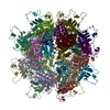
|
|---|---|
| 1 |
|
-Components
| #1: Protein | Mass: 15383.872 Da / Num. of mol.: 24 Source method: isolated from a genetically manipulated source Source: (gene. exp.) Gene: ISU1, NUA1, YPL135W / Plasmid: pET28b / Production host:  #2: Protein | Mass: 13455.976 Da / Num. of mol.: 24 Source method: isolated from a genetically manipulated source Source: (gene. exp.) Gene: YFH1, YDL120W / Plasmid: pET24a / Production host: Has protein modification | Y | |
|---|
-Experimental details
-Experiment
| Experiment | Method: ELECTRON MICROSCOPY |
|---|---|
| EM experiment | Aggregation state: PARTICLE / 3D reconstruction method: single particle reconstruction |
- Sample preparation
| Component | Name: Yfh1-Isu1 / Type: COMPLEX Details: Macromolecular complex comprising 24-mer of Yfh1 and 24-mer of Isu1 Entity ID: all / Source: RECOMBINANT | |||||||||||||||
|---|---|---|---|---|---|---|---|---|---|---|---|---|---|---|---|---|
| Molecular weight | Value: 0.7 MDa / Experimental value: NO | |||||||||||||||
| Source (natural) | Organism: | |||||||||||||||
| Source (recombinant) | Organism: | |||||||||||||||
| Buffer solution | pH: 7.3 | |||||||||||||||
| Buffer component |
| |||||||||||||||
| Specimen | Conc.: 0.11 mg/ml / Embedding applied: NO / Shadowing applied: NO / Staining applied: YES / Vitrification applied: NO Details: The protein complex was prepared by incubating Yfh1 and Isu1 (1:1.5 molar ratio) in HN100 buffer (10 mM HEPES-KOH, pH 7.3, 100 mM NaCl) and purified using Sephacryl S300 gel filtration chromatography. | |||||||||||||||
| EM staining | Type: NEGATIVE Details: Pre-incubated in HN100 buffer, the grid was placed on an 11 microliter drop of protein sample for 1 minute. Excess protein sample was blotted and washed for 3 seconds by placing the grid on ...Details: Pre-incubated in HN100 buffer, the grid was placed on an 11 microliter drop of protein sample for 1 minute. Excess protein sample was blotted and washed for 3 seconds by placing the grid on a drop of sterile water. After excess water was blotted, the grid was stained with 1% w/v uranyl acetate for 1 second and 30 seconds by successively placing it on two separate drops of uranyl acetate, with excess stain drawn off after each step. Material: uranyl acetate | |||||||||||||||
| Specimen support | Details: DV-502A instrument, Denton Vacuum Inc. / Grid material: COPPER / Grid mesh size: 400 divisions/in. / Grid type: carbon-coated, EMS |
- Electron microscopy imaging
| Experimental equipment |  Model: Tecnai F30 / Image courtesy: FEI Company |
|---|---|
| Microscopy | Model: FEI TECNAI F30 |
| Electron gun | Electron source:  FIELD EMISSION GUN / Accelerating voltage: 300 kV / Illumination mode: OTHER FIELD EMISSION GUN / Accelerating voltage: 300 kV / Illumination mode: OTHER |
| Electron lens | Mode: BRIGHT FIELD / Nominal magnification: 115000 X / Calibrated magnification: 115000 X / Nominal defocus max: 2800 nm / Nominal defocus min: 210 nm / Calibrated defocus min: 210 nm / Calibrated defocus max: 2800 nm / Cs: 2 mm / Alignment procedure: BASIC |
| Specimen holder | Cryogen: NITROGEN / Specimen holder model: SIDE ENTRY, EUCENTRIC |
| Image recording | Electron dose: 30 e/Å2 / Film or detector model: GATAN ULTRASCAN 4000 (4k x 4k) / Num. of grids imaged: 1 / Num. of real images: 475 |
| Image scans | Width: 4096 / Height: 4096 |
- Processing
| EM software |
| ||||||||||||||||||||||||||||||||||||
|---|---|---|---|---|---|---|---|---|---|---|---|---|---|---|---|---|---|---|---|---|---|---|---|---|---|---|---|---|---|---|---|---|---|---|---|---|---|
| Image processing | Details: 432 symmetry applied for reconstruction | ||||||||||||||||||||||||||||||||||||
| CTF correction | Details: The ctf.auto function from EMAN2 was applied. / Type: PHASE FLIPPING ONLY | ||||||||||||||||||||||||||||||||||||
| Particle selection | Num. of particles selected: 4218 | ||||||||||||||||||||||||||||||||||||
| Symmetry | Point symmetry: O (octahedral) | ||||||||||||||||||||||||||||||||||||
| 3D reconstruction | Resolution: 15.6 Å / Resolution method: FSC 0.143 CUT-OFF / Num. of particles: 4218 / Algorithm: FOURIER SPACE / Symmetry type: POINT | ||||||||||||||||||||||||||||||||||||
| Atomic model building | Protocol: RIGID BODY FIT / Space: REAL |
