 Movie
Movie Controller

[English] 日本語
 Yorodumi
- EMDB-8458: Zinc and the Iron Donor Frataxin Regulate Oligomerization of the ... -
+ Open data

- Basic information
| Entry | Database: EMDB / ID: EMD-8458 | |||||||||
|---|---|---|---|---|---|---|---|---|---|---|
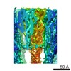
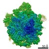
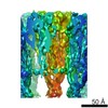
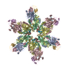
| Title | Zinc and the Iron Donor Frataxin Regulate Oligomerization of the Scaffold Protein to Form New Fe-S Cluster Assembly Centers | |||||||||
 Map data Map data | Iron sulfur cluster assembly protein 1 / Frataxin complex (generated using EMAN2) | |||||||||
 Sample Sample |
| |||||||||
 Keywords Keywords | Friedreich Ataxia / frataxin / iron-sulfur protein / mitochondria / protein complex / OXIDOREDUCTASE | |||||||||
| Function / homology |  Function and homology information Function and homology informationMitochondrial iron-sulfur cluster biogenesis / Mitochondrial protein import / Maturation of TCA enzymes and regulation of TCA cycle / Complex III assembly / mitochondrial electron transport, succinate to ubiquinone / tRNA wobble uridine modification / iron chaperone activity / iron-sulfur cluster assembly complex / heme biosynthetic process / iron-sulfur cluster assembly ...Mitochondrial iron-sulfur cluster biogenesis / Mitochondrial protein import / Maturation of TCA enzymes and regulation of TCA cycle / Complex III assembly / mitochondrial electron transport, succinate to ubiquinone / tRNA wobble uridine modification / iron chaperone activity / iron-sulfur cluster assembly complex / heme biosynthetic process / iron-sulfur cluster assembly / response to iron(II) ion / ferroxidase / ATPase activator activity / ferroxidase activity / ferric iron binding / glutathione metabolic process / iron ion transport / ferrous iron binding / mitochondrial intermembrane space / 2 iron, 2 sulfur cluster binding / response to oxidative stress / intracellular iron ion homeostasis / mitochondrial inner membrane / iron ion binding / mitochondrial matrix / mitochondrion / zinc ion binding / identical protein binding / cytoplasm Similarity search - Function | |||||||||
| Biological species |  | |||||||||
| Method | single particle reconstruction / negative staining / Resolution: 15.6 Å | |||||||||
 Authors Authors | Ranatunga W / Gakh O | |||||||||
| Funding support |  United States, 1 items United States, 1 items
| |||||||||
 Citation Citation | Journal: Metallomics / Year: 2017 Title: Zinc and the iron donor frataxin regulate oligomerization of the scaffold protein to form new Fe-S cluster assembly centers. Authors: B K Galeano / W Ranatunga / O Gakh / D Y Smith / J R Thompson / G Isaya / Abstract: Early studies of the bacterial Fe-S cluster assembly system provided structural details for how the scaffold protein and the cysteine desulfurase interact. This work and additional work on the yeast ...Early studies of the bacterial Fe-S cluster assembly system provided structural details for how the scaffold protein and the cysteine desulfurase interact. This work and additional work on the yeast and human systems elucidated a conserved mechanism for sulfur donation but did not provide any conclusive insights into the mechanism for iron delivery from the iron donor, frataxin, to the scaffold. We previously showed that oligomerization is a mechanism by which yeast frataxin (Yfh1) can promote assembly of the core machinery for Fe-S cluster synthesis both in vitro and in cells, in such a manner that the scaffold protein, Isu1, can bind to Yfh1 independent of the presence of the cysteine desulfurase, Nfs1. Here, in the absence of Yfh1, Isu1 was found to exist in two forms, one mostly monomeric with limited tendency to dimerize, and one with a strong propensity to oligomerize. Whereas the monomeric form is stabilized by zinc, the loss of zinc promotes formation of dimer and higher order oligomers. However, upon binding to oligomeric Yfh1, both forms take on a similar symmetrical trimeric configuration that places the Fe-S cluster coordinating residues of Isu1 in close proximity of iron-binding residues of Yfh1. This configuration is suitable for docking of Nfs1 in a manner that provides a structural context for coordinate iron and sulfur donation to the scaffold. Moreover, distinct structural features suggest that in physiological conditions the zinc-regulated abundance of monomeric vs. oligomeric Isu1 yields [Yfh1]·[Isu1] complexes with different Isu1 configurations that afford unique functional properties for Fe-S cluster assembly and delivery. | |||||||||
| History |
|
- Structure visualization
| Movie |
Movie viewer |
|---|---|
| Structure viewer | EM map: SurfViewMolmilJmol/JSmol |
| Supplemental images |

- Downloads & links
-EMDB archive
| Map data | emd_8458.map.gz | 19.2 MB | EMDB map data format | |
|---|---|---|---|---|
| Header (meta data) | emd-8458-v30.xmlemd-8458.xml | 14.3 KB 14.3 KB | Display Display | EMDB header |
| FSC (resolution estimation) | emd_8458_fsc.xml | 12.1 KB | Display | FSC data file |
| Images |  emd_8458.png emd_8458.png | 86.5 KB | ||
| Filedesc metadata | emd-8458.cif.gz | 6 KB | ||
| Archive directory |  http://ftp.pdbj.org/pub/emdb/structures/EMD-8458ftp://ftp.pdbj.org/pub/emdb/structures/EMD-8458 http://ftp.pdbj.org/pub/emdb/structures/EMD-8458ftp://ftp.pdbj.org/pub/emdb/structures/EMD-8458 | HTTPS FTP |
-Related structure data
| Related structure data |  5treMC M: atomic model generated by this map C: citing same article ( |
|---|---|
| Similar structure data |

-Links
| EMDB pages | EMDB (EBI/PDBe) / EMDataResource |
|---|---|
| Related items in Molecule of the Month |


-Map
| File | Download / File: emd_8458.map.gz / Format: CCP4 / Size: 91.1 MB / Type: IMAGE STORED AS FLOATING POINT NUMBER (4 BYTES) | ||||||||||||||||||||||||||||||||||||||||||||||||||||||||||||
|---|---|---|---|---|---|---|---|---|---|---|---|---|---|---|---|---|---|---|---|---|---|---|---|---|---|---|---|---|---|---|---|---|---|---|---|---|---|---|---|---|---|---|---|---|---|---|---|---|---|---|---|---|---|---|---|---|---|---|---|---|---|
| Annotation | Iron sulfur cluster assembly protein 1 / Frataxin complex (generated using EMAN2) | ||||||||||||||||||||||||||||||||||||||||||||||||||||||||||||
| Projections & slices | Image control
Images are generated by Spider. | ||||||||||||||||||||||||||||||||||||||||||||||||||||||||||||
| Voxel size | X=Y=Z: 1.034 Å | ||||||||||||||||||||||||||||||||||||||||||||||||||||||||||||
| Density |
| ||||||||||||||||||||||||||||||||||||||||||||||||||||||||||||
| Symmetry | Space group: 1 | ||||||||||||||||||||||||||||||||||||||||||||||||||||||||||||
| Details | EMDB XML:
CCP4 map header:
| ||||||||||||||||||||||||||||||||||||||||||||||||||||||||||||
 Z (Sec.)
 Y (Row.)
 X (Col.)


-Supplemental data
- Sample components
-Entire : Yfh1-Isu1
| Entire | Name: Yfh1-Isu1 |
|---|---|
| Components |
|
-Supramolecule #1: Yfh1-Isu1
| Supramolecule | Name: Yfh1-Isu1 / type: complex / ID: 1 / Parent: 0 / Macromolecule list: all Details: Macromolecular complex comprising 24-mer of Yfh1 and 24-mer of Isu1 |
|---|---|
| Source (natural) | Organism: |
| Molecular weight | Theoretical: 700 KDa |
-Macromolecule #1: Iron sulfur cluster assembly protein 1, mitochondrial
| Macromolecule | Name: Iron sulfur cluster assembly protein 1, mitochondrial / type: protein_or_peptide / ID: 1 / Number of copies: 24 / Enantiomer: LEVO |
|---|---|
| Source (natural) | Organism: |
| Molecular weight | Theoretical: 15.383872 KDa |
| Recombinant expression | Organism:  |
| Sequence | String: GSHMSSITKR LYHPKVIEHY THPRNVGSLD KKLPNVGTGL VGAPACGDVM RLQIKVNDST GVIEDVKFKT FGCGSAIASS SYMTELVQG MTLDDAAKIK NTEIAKELSL PPVKLHCSML AEDAIKAAIK DYKSKRNTPT MLS UniProtKB: Iron sulfur cluster assembly protein 1, mitochondrial |
-Macromolecule #2: Frataxin homolog, mitochondrial
| Macromolecule | Name: Frataxin homolog, mitochondrial / type: protein_or_peptide / ID: 2 / Number of copies: 24 / Enantiomer: LEVO / EC number: ferroxidase |
|---|---|
| Source (natural) | Organism: |
| Molecular weight | Theoretical: 13.455976 KDa |
| Recombinant expression | Organism: |
| Sequence | String: VESSTDGQVV PQEVLNLPLE KAHEEADDYL DHLLDSLEEL SEAHPDCIPD VELSHGVMTL EIPAFGTYVI NKQPPNKQIW LASPLSGPN RFDLLNGEWV SLRNGTKLTD ILTEEVEKAI SK UniProtKB: Frataxin homolog, mitochondrial |
-Experimental details
-Structure determination
| Method | negative staining |
|---|---|
 Processing Processing | single particle reconstruction |
| Aggregation state | particle |
-Sample preparation
| Concentration | 0.11 mg/mL | |||||||||
|---|---|---|---|---|---|---|---|---|---|---|
| Buffer | pH: 7.3 Component:
| |||||||||
| Staining | Type: NEGATIVE / Material: uranyl acetate Details: Pre-incubated in HN100 buffer, the grid was placed on an 11 microliter drop of protein sample for 1 minute. Excess protein sample was blotted and washed for 3 seconds by placing the grid on ...Details: Pre-incubated in HN100 buffer, the grid was placed on an 11 microliter drop of protein sample for 1 minute. Excess protein sample was blotted and washed for 3 seconds by placing the grid on a drop of sterile water. After excess water was blotted, the grid was stained with 1% w/v uranyl acetate for 1 second and 30 seconds by successively placing it on two separate drops of uranyl acetate, with excess stain drawn off after each step. | |||||||||
| Grid | Model: carbon-coated, EMS / Material: COPPER / Mesh: 400 / Pretreatment - Type: GLOW DISCHARGE / Pretreatment - Time: 30 sec. / Details: DV-502A instrument, Denton Vacuum Inc. | |||||||||
| Details | The protein complex was prepared by incubating Yfh1 and Isu1 (1:1.5 molar ratio) in HN100 buffer (10 mM HEPES-KOH, pH 7.3, 100 mM NaCl) and purified using Sephacryl S300 gel filtration chromatography. |
- Electron microscopy
| Microscope | FEI TECNAI F30 |
|---|---|
| Image recording | Film or detector model: GATAN ULTRASCAN 4000 (4k x 4k) / Digitization - Dimensions - Width: 4096 pixel / Digitization - Dimensions - Height: 4096 pixel / Number grids imaged: 1 / Number real images: 475 / Average electron dose: 30.0 e/Å2 |
| Electron beam | Acceleration voltage: 300 kV / Electron source:  FIELD EMISSION GUN FIELD EMISSION GUN |
| Electron optics | Calibrated defocus max: 2.8000000000000003 µm / Calibrated defocus min: 0.21 µm / Calibrated magnification: 115000 / Illumination mode: OTHER / Imaging mode: BRIGHT FIELD / Cs: 2.0 mm / Nominal defocus max: 2.8000000000000003 µm / Nominal defocus min: 0.21 µm / Nominal magnification: 115000 |
| Sample stage | Specimen holder model: SIDE ENTRY, EUCENTRIC / Cooling holder cryogen: NITROGEN |
| Experimental equipment |  Model: Tecnai F30 / Image courtesy: FEI Company |
+Image processing

-Atomic model buiding 1
| Refinement | Space: REAL / Protocol: RIGID BODY FIT |
|---|---|
| Output model | PDB-5tre: |
