 Movie
Movie Controller

[English] 日本語
 Yorodumi
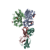
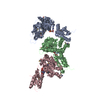
- PDB-5tq0: Crystal structure of amino terminal domains of the NMDA receptor ... -
+ Open data

- Basic information
| Entry | Database: PDB / ID: 5tq0 | |||||||||
|---|---|---|---|---|---|---|---|---|---|---|
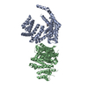
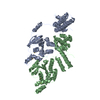
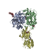
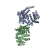
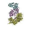
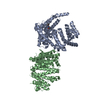
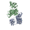
| Title | Crystal structure of amino terminal domains of the NMDA receptor subunit GluN1 and GluN2A in the presence of EDTA | |||||||||
 Components Components |
| |||||||||
 Keywords Keywords | TRANSPORT PROTEIN / ION CHANNEL / NMDA RECEPTOR / ALLOSTERIC MODULATION / ZINC INHIBITION / IMMUNE SYSTEM | |||||||||
| Function / homology |  Function and homology information Function and homology informationneurotransmitter receptor transport, plasma membrane to endosome / regulation of response to alcohol / response to ammonium ion / receptor recycling / directional locomotion / response to environmental enrichment / Assembly and cell surface presentation of NMDA receptors / response to hydrogen sulfide / auditory behavior / serotonin metabolic process ...neurotransmitter receptor transport, plasma membrane to endosome / regulation of response to alcohol / response to ammonium ion / receptor recycling / directional locomotion / response to environmental enrichment / Assembly and cell surface presentation of NMDA receptors / response to hydrogen sulfide / auditory behavior / serotonin metabolic process / response to other organism / protein localization to postsynaptic membrane / regulation of ARF protein signal transduction / cellular response to magnesium ion / positive regulation of inhibitory postsynaptic potential / response to methylmercury / response to manganese ion / response to carbohydrate / sleep / regulation of NMDA receptor activity / locomotion / dendritic spine organization / cellular response to dsRNA / cellular response to lipid / regulation of monoatomic cation transmembrane transport / NMDA glutamate receptor activity / Synaptic adhesion-like molecules / voltage-gated monoatomic cation channel activity / response to glycoside / NMDA selective glutamate receptor complex / glutamate binding / ligand-gated sodium channel activity / glutamate receptor signaling pathway / calcium ion transmembrane import into cytosol / response to zinc ion / spinal cord development / response to amine / parallel fiber to Purkinje cell synapse / cellular response to zinc ion / startle response / monoatomic cation transmembrane transport / dopamine metabolic process / response to lithium ion / response to light stimulus / regulation of postsynaptic membrane potential / cellular response to glycine / modulation of excitatory postsynaptic potential / action potential / conditioned place preference / regulation of neuronal synaptic plasticity / positive regulation of protein targeting to membrane / glutamate receptor binding / positive regulation of excitatory postsynaptic potential / Unblocking of NMDA receptors, glutamate binding and activation / neuron development / positive regulation of synaptic transmission, glutamatergic / multicellular organismal response to stress / postsynaptic density, intracellular component / monoatomic cation channel activity / response to fungicide / glutamate-gated receptor activity / cell adhesion molecule binding / cellular response to manganese ion / glutamate-gated calcium ion channel activity / neurogenesis / sensory perception of pain / dendrite membrane / ligand-gated monoatomic ion channel activity involved in regulation of presynaptic membrane potential / sodium ion transmembrane transport / ionotropic glutamate receptor signaling pathway / protein tyrosine kinase binding / cytoplasmic vesicle membrane / synaptic membrane / response to amphetamine / learning / regulation of membrane potential / excitatory postsynaptic potential / response to nicotine / response to cocaine / transmitter-gated monoatomic ion channel activity involved in regulation of postsynaptic membrane potential / hippocampus development / synaptic transmission, glutamatergic / cellular response to amino acid stimulus / protein catabolic process / regulation of long-term neuronal synaptic plasticity / response to calcium ion / postsynaptic density membrane / cerebral cortex development / negative regulation of protein catabolic process / visual learning / regulation of synaptic plasticity / modulation of chemical synaptic transmission / calcium ion transmembrane transport / cellular response to growth factor stimulus / calcium channel activity / response to wounding / response to lead ion / memory / long-term synaptic potentiation / terminal bouton Similarity search - Function | |||||||||
| Biological species |  | |||||||||
| Method |  X-RAY DIFFRACTION / SYNCHROTRON / MOLECULAR REPLACEMENT / Resolution: 2.7 Å X-RAY DIFFRACTION / SYNCHROTRON / MOLECULAR REPLACEMENT / Resolution: 2.7 Å | |||||||||
 Authors Authors | Romero-Hernandez, A. / Simorowski, N. / Karakas, E. / Furukawa, H. | |||||||||
| Funding support |  United States, 2items United States, 2items
| |||||||||
 Citation Citation | Journal: Neuron / Year: 2016 Title: Molecular Basis for Subtype Specificity and High-Affinity Zinc Inhibition in the GluN1-GluN2A NMDA Receptor Amino-Terminal Domain. Authors: Romero-Hernandez, A. / Simorowski, N. / Karakas, E. / Furukawa, H. | |||||||||
| History |
|
- Structure visualization
| Structure viewer | Molecule: MolmilJmol/JSmol |
|---|
- Downloads & links
-Download
| PDBx/mmCIF format | 5tq0.cif.gz | 231.4 KB | Display | PDBx/mmCIF format |
|---|---|---|---|---|
| PDB format | pdb5tq0.ent.gz | 176.6 KB | Display | PDB format |
| PDBx/mmJSON format | 5tq0.json.gz | Tree view | PDBx/mmJSON format | |
| Others |  Other downloads Other downloads |
-Validation report
| Summary document | 5tq0_validation.pdf.gz | 512.9 KB | Display | wwPDB validaton report |
|---|---|---|---|---|
| Full document | 5tq0_full_validation.pdf.gz | 525.3 KB | Display | |
| Data in XML | 5tq0_validation.xml.gz | 44.9 KB | Display | |
| Data in CIF | 5tq0_validation.cif.gz | 58.3 KB | Display | |
| Arichive directory | https://data.pdbj.org/pub/pdb/validation_reports/tq/5tq0ftp://data.pdbj.org/pub/pdb/validation_reports/tq/5tq0 | HTTPS FTP |
-Related structure data
| Related structure data | 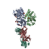 5tpwSC 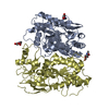 5tpzC  5tq2C C: citing same article ( S: Starting model for refinement |
|---|---|
| Similar structure data |

-Links
 PDBj
PDBj


- Assembly
| Deposited unit | 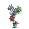
| ||||||||
|---|---|---|---|---|---|---|---|---|---|
| 1 |
| ||||||||
| Unit cell |
|
-Components
-Protein , 2 types, 2 molecules AB
| #1: Protein | Mass: 43801.043 Da / Num. of mol.: 1 / Fragment: residues 24-408 / Mutation: N38Q, N348Q Source method: isolated from a genetically manipulated source Source: (gene. exp.)  Trichoplusia ni (cabbage looper) / References: UniProt: Q91977, UniProt: A0A1L8F5J9*PLUS Trichoplusia ni (cabbage looper) / References: UniProt: Q91977, UniProt: A0A1L8F5J9*PLUS |
|---|---|
| #2: Protein | Mass: 40572.078 Da / Num. of mol.: 1 / Fragment: residues 34-393 Source method: isolated from a genetically manipulated source Source: (gene. exp.) Trichoplusia ni (cabbage looper) / References: UniProt: Q00959 |
-Antibody , 2 types, 2 molecules HL
| #3: Antibody | Mass: 23993.883 Da / Num. of mol.: 1 / Source method: isolated from a natural source / Source: (natural) |
|---|---|
| #4: Antibody | Mass: 23568.025 Da / Num. of mol.: 1 / Source method: isolated from a natural source / Source: (natural) |
-Sugars , 1 types, 2 molecules 
| #5: Sugar |  Type: D-saccharide, beta linking / Mass: 221.208 Da / Num. of mol.: 2 Type: D-saccharide, beta linking / Mass: 221.208 Da / Num. of mol.: 2Source method: isolated from a genetically manipulated source Formula: C8H15NO6 |
|---|
-Non-polymers , 4 types, 65 molecules 


| #6: Chemical |  Mass: 92.094 Da / Num. of mol.: 2 / Source method: obtained synthetically / Formula: C3H8O3 Mass: 92.094 Da / Num. of mol.: 2 / Source method: obtained synthetically / Formula: C3H8O3#7: Chemical | ChemComp-SO4 /  Mass: 96.063 Da / Num. of mol.: 12 / Source method: obtained synthetically / Formula: SO4 Mass: 96.063 Da / Num. of mol.: 12 / Source method: obtained synthetically / Formula: SO4#8: Chemical | ChemComp-IPA / |  Mass: 60.095 Da / Num. of mol.: 1 / Source method: obtained synthetically / Formula: C3H8O Mass: 60.095 Da / Num. of mol.: 1 / Source method: obtained synthetically / Formula: C3H8O#9: Water | ChemComp-HOH / | Mass: 18.015 Da / Num. of mol.: 50 / Source method: isolated from a natural source / Formula: H2O |
|---|
-Details
| Has protein modification | Y |
|---|
-Experimental details
-Experiment
| Experiment | Method: X-RAY DIFFRACTION / Number of used crystals: 1 |
|---|
- Sample preparation
| Crystal | Density Matthews: 2.81 Å3/Da / Density % sol: 56.19 % |
|---|---|
| Crystal grow | Temperature: 291 K / Method: vapor diffusion, hanging drop / Details: 1.8 M Ammonium sulfate, 2.5% Isopropanol |
-Data collection
| Diffraction | Mean temperature: 100 K |
|---|---|
| Diffraction source | Source: SYNCHROTRON / Site: APS / Beamline: 23-ID-D / Wavelength: 1.28 Å |
| Detector | Type: DECTRIS PILATUS3 S 6M / Detector: PIXEL / Date: Apr 8, 2015 |
| Radiation | Protocol: SINGLE WAVELENGTH / Monochromatic (M) / Laue (L): M / Scattering type: x-ray |
| Radiation wavelength | Wavelength: 1.28 Å / Relative weight: 1 |
| Reflection | Resolution: 2.7→50 Å / Num. obs: 77929 / % possible obs: 99.2 % / Redundancy: 3.1 % / Rmerge(I) obs: 0.129 / Net I/σ(I): 7.72 |
- Processing
| Software |
| |||||||||||||||||||||||||||||||||||||||||||||||||||||||||||||||||||||||||||||||||||||||||||||||||||||||||||||||||||||||||||||||||||||||||||||||||||||||||||||||||||||||||||||||||||||||||||||||||||||||||||||||||||||||||
|---|---|---|---|---|---|---|---|---|---|---|---|---|---|---|---|---|---|---|---|---|---|---|---|---|---|---|---|---|---|---|---|---|---|---|---|---|---|---|---|---|---|---|---|---|---|---|---|---|---|---|---|---|---|---|---|---|---|---|---|---|---|---|---|---|---|---|---|---|---|---|---|---|---|---|---|---|---|---|---|---|---|---|---|---|---|---|---|---|---|---|---|---|---|---|---|---|---|---|---|---|---|---|---|---|---|---|---|---|---|---|---|---|---|---|---|---|---|---|---|---|---|---|---|---|---|---|---|---|---|---|---|---|---|---|---|---|---|---|---|---|---|---|---|---|---|---|---|---|---|---|---|---|---|---|---|---|---|---|---|---|---|---|---|---|---|---|---|---|---|---|---|---|---|---|---|---|---|---|---|---|---|---|---|---|---|---|---|---|---|---|---|---|---|---|---|---|---|---|---|---|---|---|---|---|---|---|---|---|---|---|---|---|---|---|---|---|---|---|
| Refinement | Method to determine structure: MOLECULAR REPLACEMENT Starting model: 5TPW Resolution: 2.7→47.569 Å / SU ML: 0.39 / Cross valid method: FREE R-VALUE / σ(F): 1.33 / Phase error: 25.61 / Stereochemistry target values: ML
| |||||||||||||||||||||||||||||||||||||||||||||||||||||||||||||||||||||||||||||||||||||||||||||||||||||||||||||||||||||||||||||||||||||||||||||||||||||||||||||||||||||||||||||||||||||||||||||||||||||||||||||||||||||||||
| Solvent computation | Shrinkage radii: 0.9 Å / VDW probe radii: 1.11 Å / Solvent model: FLAT BULK SOLVENT MODEL | |||||||||||||||||||||||||||||||||||||||||||||||||||||||||||||||||||||||||||||||||||||||||||||||||||||||||||||||||||||||||||||||||||||||||||||||||||||||||||||||||||||||||||||||||||||||||||||||||||||||||||||||||||||||||
| Displacement parameters | Biso max: 147.64 Å2 / Biso mean: 66.2492 Å2 / Biso min: 28.67 Å2 | |||||||||||||||||||||||||||||||||||||||||||||||||||||||||||||||||||||||||||||||||||||||||||||||||||||||||||||||||||||||||||||||||||||||||||||||||||||||||||||||||||||||||||||||||||||||||||||||||||||||||||||||||||||||||
| Refinement step | Cycle: final / Resolution: 2.7→47.569 Å
| |||||||||||||||||||||||||||||||||||||||||||||||||||||||||||||||||||||||||||||||||||||||||||||||||||||||||||||||||||||||||||||||||||||||||||||||||||||||||||||||||||||||||||||||||||||||||||||||||||||||||||||||||||||||||
| Refine LS restraints |
| |||||||||||||||||||||||||||||||||||||||||||||||||||||||||||||||||||||||||||||||||||||||||||||||||||||||||||||||||||||||||||||||||||||||||||||||||||||||||||||||||||||||||||||||||||||||||||||||||||||||||||||||||||||||||
| LS refinement shell | Refine-ID: X-RAY DIFFRACTION / Total num. of bins used: 30
|
