 Movie
Movie Controller

[English] 日本語
 Yorodumi
- PDB-5tpz: Crystal structure of amino terminal domains of the NMDA receptor ... -
+ Open data

- Basic information
| Entry | Database: PDB / ID: 5tpz | |||||||||
|---|---|---|---|---|---|---|---|---|---|---|
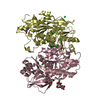
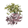
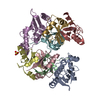
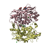
| Title | Crystal structure of amino terminal domains of the NMDA receptor subunit GluN1 and GluN2B in apo closed state | |||||||||
 Components Components |
| |||||||||
 Keywords Keywords | TRANSPORT PROTEIN / ION CHANNEL / NMDA RECEPTOR / ALLOSTERIC MODULATION | |||||||||
| Function / homology |  Function and homology information Function and homology informationcellular response to corticosterone stimulus / cellular response to magnesium starvation / sensory organ development / cellular response to curcumin / regulation of cAMP/PKA signal transduction / EPHB-mediated forward signaling / Assembly and cell surface presentation of NMDA receptors / auditory behavior / sensitization / response to other organism ...cellular response to corticosterone stimulus / cellular response to magnesium starvation / sensory organ development / cellular response to curcumin / regulation of cAMP/PKA signal transduction / EPHB-mediated forward signaling / Assembly and cell surface presentation of NMDA receptors / auditory behavior / sensitization / response to other organism / response to hydrogen sulfide / dendritic branch / fear response / response to methylmercury / regulation of ARF protein signal transduction / apical dendrite / response to manganese ion / suckling behavior / interleukin-1 receptor binding / response to carbohydrate / cellular response to lipid / cellular response to dsRNA / RAF/MAP kinase cascade / negative regulation of dendritic spine maintenance / positive regulation of inhibitory postsynaptic potential / response to amine / response to growth hormone / heterocyclic compound binding / Synaptic adhesion-like molecules / response to glycoside / regulation of monoatomic cation transmembrane transport / NMDA glutamate receptor activity / NMDA selective glutamate receptor complex / glutamate binding / response to zinc ion / ligand-gated sodium channel activity / calcium ion transmembrane import into cytosol / positive regulation of glutamate secretion / protein heterotetramerization / small molecule binding / glycine binding / receptor clustering / startle response / parallel fiber to Purkinje cell synapse / regulation of MAPK cascade / regulation of postsynaptic membrane potential / behavioral response to pain / response to electrical stimulus / monoatomic cation transmembrane transport / extracellularly glutamate-gated ion channel activity / associative learning / action potential / response to magnesium ion / Unblocking of NMDA receptors, glutamate binding and activation / regulation of neuronal synaptic plasticity / monoatomic cation transport / glutamate receptor binding / detection of mechanical stimulus involved in sensory perception of pain / ligand-gated monoatomic ion channel activity / multicellular organismal response to stress / response to mechanical stimulus / neuron development / long-term memory / postsynaptic density, intracellular component / behavioral fear response / monoatomic cation channel activity / synaptic cleft / response to fungicide / cellular response to manganese ion / glutamate-gated receptor activity / regulation of long-term synaptic depression / positive regulation of synaptic transmission, glutamatergic / glutamate-gated calcium ion channel activity / presynaptic active zone membrane / response to cytokine / D2 dopamine receptor binding / cell adhesion molecule binding / ionotropic glutamate receptor signaling pathway / ionotropic glutamate receptor binding / ligand-gated monoatomic ion channel activity involved in regulation of presynaptic membrane potential / protein tyrosine kinase binding / cellular response to forskolin / positive regulation of excitatory postsynaptic potential / hippocampal mossy fiber to CA3 synapse / protein serine/threonine kinase binding / sodium ion transmembrane transport / learning / response to nicotine / synaptic membrane / response to amphetamine / hippocampus development / response to cocaine / cellular response to amino acid stimulus / transmitter-gated monoatomic ion channel activity involved in regulation of postsynaptic membrane potential / synaptic transmission, glutamatergic / regulation of membrane potential / excitatory postsynaptic potential / cerebral cortex development / response to calcium ion / regulation of synaptic plasticity Similarity search - Function | |||||||||
| Biological species |  | |||||||||
| Method |  X-RAY DIFFRACTION / SYNCHROTRON / MOLECULAR REPLACEMENT / Resolution: 3.095 Å X-RAY DIFFRACTION / SYNCHROTRON / MOLECULAR REPLACEMENT / Resolution: 3.095 Å | |||||||||
 Authors Authors | Romero-Hernandez, A. / Simorwski, N. / Karakas, E. / Furukawa, H. | |||||||||
| Funding support |  United States, 2items United States, 2items
| |||||||||
 Citation Citation | Journal: Neuron / Year: 2016 Title: Molecular Basis for Subtype Specificity and High-Affinity Zinc Inhibition in the GluN1-GluN2A NMDA Receptor Amino-Terminal Domain. Authors: Romero-Hernandez, A. / Simorowski, N. / Karakas, E. / Furukawa, H. | |||||||||
| History |
|
- Structure visualization
| Structure viewer | Molecule: MolmilJmol/JSmol |
|---|
- Downloads & links
-Download
| PDBx/mmCIF format | 5tpz.cif.gz | 148.4 KB | Display | PDBx/mmCIF format |
|---|---|---|---|---|
| PDB format | pdb5tpz.ent.gz | 108 KB | Display | PDB format |
| PDBx/mmJSON format | 5tpz.json.gz | Tree view | PDBx/mmJSON format | |
| Others |  Other downloads Other downloads |
-Validation report
| Arichive directory | https://data.pdbj.org/pub/pdb/validation_reports/tp/5tpzftp://data.pdbj.org/pub/pdb/validation_reports/tp/5tpz | HTTPS FTP |
|---|
-Related structure data
| Related structure data | 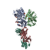 5tpwC 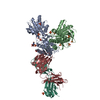 5tq0C 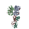 5tq2C 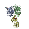 3qelS S: Starting model for refinement C: citing same article ( |
|---|---|
| Similar structure data |

-Links
 PDBj
PDBj


- Assembly
| Deposited unit | 
| ||||||||
|---|---|---|---|---|---|---|---|---|---|
| 1 |
| ||||||||
| Unit cell |
| ||||||||
| Components on special symmetry positions |
|
-Components
| #1: Protein | Mass: 42932.055 Da / Num. of mol.: 1 / Fragment: residues 23-405 Source method: isolated from a genetically manipulated source Source: (gene. exp.)  Trichoplusia ni (cabbage looper) / References: UniProt: Q91977, UniProt: A0A1L8F5J9*PLUS Trichoplusia ni (cabbage looper) / References: UniProt: Q91977, UniProt: A0A1L8F5J9*PLUS | ||||
|---|---|---|---|---|---|
| #2: Protein | Mass: 41149.625 Da / Num. of mol.: 1 / Fragment: residues 32-393 Source method: isolated from a genetically manipulated source Source: (gene. exp.) Trichoplusia ni (cabbage looper) / References: UniProt: Q00960 | ||||
| #3: Sugar | ChemComp-NAG /   Type: D-saccharide, beta linking / Mass: 221.208 Da / Num. of mol.: 4 Type: D-saccharide, beta linking / Mass: 221.208 Da / Num. of mol.: 4Source method: isolated from a genetically manipulated source Formula: C8H15NO6 #4: Water | ChemComp-HOH / |  Mass: 18.015 Da / Num. of mol.: 4 / Source method: isolated from a natural source / Formula: H2O Mass: 18.015 Da / Num. of mol.: 4 / Source method: isolated from a natural source / Formula: H2OHas protein modification | Y | |
-Experimental details
-Experiment
| Experiment | Method: X-RAY DIFFRACTION / Number of used crystals: 1 |
|---|
- Sample preparation
| Crystal | Density Matthews: 2.7 Å3/Da / Density % sol: 54.51 % |
|---|---|
| Crystal grow | Temperature: 291 K / Method: vapor diffusion, hanging drop / pH: 5 Details: 20% PEG 3350, 1.4M Sodium formate, 0.3M Sodium thyocyanate, 0.1M sodium acetate pH 5.0 |
-Data collection
| Diffraction | Mean temperature: 100 K |
|---|---|
| Diffraction source | Source: SYNCHROTRON / Site: NSLS / Beamline: X25 / Wavelength: 1 Å |
| Detector | Type: ADSC QUANTUM 315 / Detector: CCD / Date: Jun 12, 2010 |
| Radiation | Protocol: SINGLE WAVELENGTH / Monochromatic (M) / Laue (L): M / Scattering type: x-ray |
| Radiation wavelength | Wavelength: 1 Å / Relative weight: 1 |
| Reflection | Resolution: 3.09→37.891 Å / Num. obs: 18198 / % possible obs: 98.6 % / Redundancy: 5.2 % / Rmerge(I) obs: 0.093 / Net I/σ(I): 13 |
- Processing
| Software |
| ||||||||||||||||||||||||||||||||||||||||||||||||||||||||
|---|---|---|---|---|---|---|---|---|---|---|---|---|---|---|---|---|---|---|---|---|---|---|---|---|---|---|---|---|---|---|---|---|---|---|---|---|---|---|---|---|---|---|---|---|---|---|---|---|---|---|---|---|---|---|---|---|---|
| Refinement | Method to determine structure: MOLECULAR REPLACEMENT Starting model: 3QEL Resolution: 3.095→37.891 Å / SU ML: 0.5 / Cross valid method: FREE R-VALUE / σ(F): 1.34 / Phase error: 33.42
| ||||||||||||||||||||||||||||||||||||||||||||||||||||||||
| Solvent computation | Shrinkage radii: 0.9 Å / VDW probe radii: 1.11 Å | ||||||||||||||||||||||||||||||||||||||||||||||||||||||||
| Displacement parameters | Biso max: 149.19 Å2 / Biso mean: 86.3075 Å2 / Biso min: 48.34 Å2 | ||||||||||||||||||||||||||||||||||||||||||||||||||||||||
| Refinement step | Cycle: final / Resolution: 3.095→37.891 Å
| ||||||||||||||||||||||||||||||||||||||||||||||||||||||||
| Refine LS restraints |
| ||||||||||||||||||||||||||||||||||||||||||||||||||||||||
| LS refinement shell | Refine-ID: X-RAY DIFFRACTION / Total num. of bins used: 7
|
