National Institutes of Health/National Institute of General Medical Sciences (NIH/NIGMS)
R01GM085234
United States
National Institutes of Health/National Institute of Neurological Disorders and Stroke (NIH/NINDS)
RO1NS053494
United States
National Basic Research Program of China (973 Program)
2014CB910301
China
National Natural Science Foundation of China (NSFC)
31370821
China
Top Talents Program of Yunnan Province
2011HA012
China
High-level Overseas Talents of Yunnan Province
China
China Youth 1000-Talent Program of the State Council of China
China
Beijing Advanced Innovation Center for Structural Biology
China
Citation
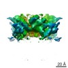
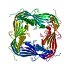
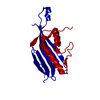
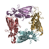
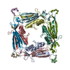
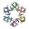
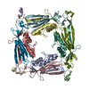
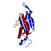
Journal: Nat Struct Mol Biol / Year: 2017 Title: Structural basis of dual Ca/pH regulation of the endolysosomal TRPML1 channel. Authors: Minghui Li / Wei K Zhang / Nicole M Benvin / Xiaoyuan Zhou / Deyuan Su / Huan Li / Shu Wang / Ioannis E Michailidis / Liang Tong / Xueming Li / Jian Yang / Abstract: The activities of organellar ion channels are often regulated by Ca and H, which are present in high concentrations in many organelles. Here we report a structural element critical for dual Ca/pH ...The activities of organellar ion channels are often regulated by Ca and H, which are present in high concentrations in many organelles. Here we report a structural element critical for dual Ca/pH regulation of TRPML1, a Ca-release channel crucial for endolysosomal function. TRPML1 mutations cause mucolipidosis type IV (MLIV), a severe lysosomal storage disorder characterized by neurodegeneration, mental retardation and blindness. We obtained crystal structures of the 213-residue luminal domain of human TRPML1 containing three missense MLIV-causing mutations. This domain forms a tetramer with a highly electronegative central pore formed by a novel luminal pore loop. Cysteine cross-linking and cryo-EM analyses confirmed that this architecture occurs in the full-length channel. Structure-function studies demonstrated that Ca and H interact with the luminal pore and exert physiologically important regulation. The MLIV-causing mutations disrupt the luminal-domain structure and cause TRPML1 mislocalization. Our study reveals the structural underpinnings of TRPML1's regulation, assembly and pathogenesis.
In the structure databanks used in Yorodumi, some data are registered as the other names, "COVID-19 virus" and "2019-nCoV". Here are the details of the virus and the list of structure data.
Jan 31, 2019. EMDB accession codes are about to change! (news from PDBe EMDB page)
EMDB accession codes are about to change! (news from PDBe EMDB page)
The allocation of 4 digits for EMDB accession codes will soon come to an end. Whilst these codes will remain in use, new EMDB accession codes will include an additional digit and will expand incrementally as the available range of codes is exhausted. The current 4-digit format prefixed with “EMD-” (i.e. EMD-XXXX) will advance to a 5-digit format (i.e. EMD-XXXXX), and so on. It is currently estimated that the 4-digit codes will be depleted around Spring 2019, at which point the 5-digit format will come into force.
The EM Navigator/Yorodumi systems omit the EMD- prefix.
Related info.:Q: What is EMD? / ID/Accession-code notation in Yorodumi/EM Navigator
Yorodumi is a browser for structure data from EMDB, PDB, SASBDB, etc.
This page is also the successor to EM Navigator detail page, and also detail information page/front-end page for Omokage search.
The word "yorodu" (or yorozu) is an old Japanese word meaning "ten thousand". "mi" (miru) is to see.
Related info.:EMDB / PDB / SASBDB / Comparison of 3 databanks / Yorodumi Search / Aug 31, 2016. New EM Navigator & Yorodumi / Yorodumi Papers / Jmol/JSmol / Function and homology information / Changes in new EM Navigator and Yorodumi
 Movie
Movie Controller


 Open data
 Basic information
 Components
 Keywords
 Function and homology information
 Homo sapiens (human)
 X-RAY DIFFRACTION /
  Authors
 United States,
  China, 8items
  Citation
 Structure visualization
 Downloads & links
 Other downloads
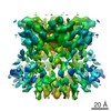


 PDBj
PDBj
 Assembly
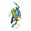

 Mass: 18.015 Da / Num. of mol.: 66 / Source method: isolated from a natural source / Formula: H2O
 Sample preparation
 Processing