 Movie
Movie Controller

[English] 日本語
 Yorodumi
- PDB-5t9b: Crystal structure of B. subtilis 168 GlpQ in complex with glycero... -
+ Open data

- Basic information
| Entry | Database: PDB / ID: 5t9b | |||||||||
|---|---|---|---|---|---|---|---|---|---|---|
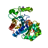
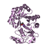
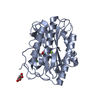
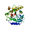
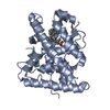
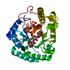
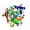
| Title | Crystal structure of B. subtilis 168 GlpQ in complex with glycerol-3-phosphate (5 minute soak) | |||||||||
 Components Components | Glycerophosphoryl diester phosphodiesterase | |||||||||
 Keywords Keywords | HYDROLASE / metal binding protein | |||||||||
| Function / homology |  Function and homology information Function and homology informationglycerophosphodiester phosphodiesterase / glycerophosphodiester phosphodiesterase activity / glycerol metabolic process / phosphoric diester hydrolase activity / lipid metabolic process / cell wall organization / extracellular region / metal ion binding Similarity search - Function | |||||||||
| Biological species |  | |||||||||
| Method |  X-RAY DIFFRACTION / SYNCHROTRON / MOLECULAR REPLACEMENT / Resolution: 1.62 Å X-RAY DIFFRACTION / SYNCHROTRON / MOLECULAR REPLACEMENT / Resolution: 1.62 Å | |||||||||
 Authors Authors | Li, F.K.K. / Strynadka, N.C.J. | |||||||||
| Funding support |  Canada, Canada,  United States, 2items United States, 2items
| |||||||||
 Citation Citation | Journal: J. Biol. Chem. / Year: 2016 Title: Identification of Two Phosphate Starvation-induced Wall Teichoic Acid Hydrolases Provides First Insights into the Degradative Pathway of a Key Bacterial Cell Wall Component. Authors: Myers, C.L. / Li, F.K. / Koo, B.M. / El-Halfawy, O.M. / French, S. / Gross, C.A. / Strynadka, N.C. / Brown, E.D. | |||||||||
| History |
|
- Structure visualization
| Structure viewer | Molecule: MolmilJmol/JSmol |
|---|
- Downloads & links
-Download
| PDBx/mmCIF format | 5t9b.cif.gz | 126.8 KB | Display | PDBx/mmCIF format |
|---|---|---|---|---|
| PDB format | pdb5t9b.ent.gz | 93.9 KB | Display | PDB format |
| PDBx/mmJSON format | 5t9b.json.gz | Tree view | PDBx/mmJSON format | |
| Others |  Other downloads Other downloads |
-Validation report
| Arichive directory | https://data.pdbj.org/pub/pdb/validation_reports/t9/5t9bftp://data.pdbj.org/pub/pdb/validation_reports/t9/5t9b | HTTPS FTP |
|---|
-Related structure data
| Related structure data |  5t91C  5t9cC  4r70S C: citing same article ( S: Starting model for refinement |
|---|---|
| Similar structure data |


-Links
 PDBj
PDBj- Assembly
| Deposited unit | 
| ||||||||||||
|---|---|---|---|---|---|---|---|---|---|---|---|---|---|
| 1 |
| ||||||||||||
| Unit cell |
|
-Components
| #1: Protein | Mass: 30202.641 Da / Num. of mol.: 1 / Fragment: UNP residues 27-293 Source method: isolated from a genetically manipulated source Source: (gene. exp.) References: UniProt: P37965, glycerophosphodiester phosphodiesterase |
|---|---|
| #2: Chemical | ChemComp-CA /   Mass: 40.078 Da / Num. of mol.: 1 / Source method: obtained synthetically / Formula: Ca Mass: 40.078 Da / Num. of mol.: 1 / Source method: obtained synthetically / Formula: Ca |
| #3: Chemical | ChemComp-G3P /   Mass: 172.074 Da / Num. of mol.: 1 / Source method: obtained synthetically / Formula: C3H9O6P Mass: 172.074 Da / Num. of mol.: 1 / Source method: obtained synthetically / Formula: C3H9O6P |
| #4: Chemical | ChemComp-NA /   Mass: 22.990 Da / Num. of mol.: 1 / Source method: obtained synthetically / Formula: Na Mass: 22.990 Da / Num. of mol.: 1 / Source method: obtained synthetically / Formula: Na |
| #5: Water | ChemComp-HOH /  Mass: 18.015 Da / Num. of mol.: 193 / Source method: isolated from a natural source / Formula: H2O Mass: 18.015 Da / Num. of mol.: 193 / Source method: isolated from a natural source / Formula: H2O |
-Experimental details
-Experiment
| Experiment | Method: X-RAY DIFFRACTION / Number of used crystals: 1 |
|---|
- Sample preparation
| Crystal | Density Matthews: 2.22 Å3/Da / Density % sol: 44.7 % |
|---|---|
| Crystal grow | Temperature: 298 K / Method: vapor diffusion, sitting drop / pH: 8.5 / Details: 100 mM bicine, pH 8.5, 25% PEG6000 |
-Data collection
| Diffraction | Mean temperature: 100 K |
|---|---|
| Diffraction source | Source: SYNCHROTRON / Site: CLSI / Beamline: 08ID-1 / Wavelength: 0.979 Å |
| Detector | Type: RAYONIX MX-300 / Detector: CCD / Date: Mar 9, 2016 |
| Radiation | Monochromator: double crystal Si(111) / Protocol: SINGLE WAVELENGTH / Monochromatic (M) / Laue (L): M / Scattering type: x-ray |
| Radiation wavelength | Wavelength: 0.979 Å / Relative weight: 1 |
| Reflection | Resolution: 1.62→43.83 Å / Num. obs: 34957 / % possible obs: 99.92 % / Redundancy: 6.7 % / CC1/2: 0.999 / Net I/σ(I): 19.4 |
| Reflection shell | Resolution: 1.62→1.68 Å / Redundancy: 6.7 % / Mean I/σ(I) obs: 1.6 / CC1/2: 0.854 / % possible all: 99.91 |
- Processing
| Software |
| ||||||||||||||||
|---|---|---|---|---|---|---|---|---|---|---|---|---|---|---|---|---|---|
| Refinement | Method to determine structure: MOLECULAR REPLACEMENT Starting model: PDB entry 4R70 Resolution: 1.62→43.83 Å / Cross valid method: THROUGHOUT
| ||||||||||||||||
| Refinement step | Cycle: LAST / Resolution: 1.62→43.83 Å
| ||||||||||||||||
| LS refinement shell | Resolution: 1.62→1.68 Å
|
