 Movie
Movie Controller

[English] 日本語
 Yorodumi
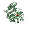
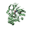
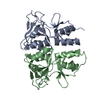
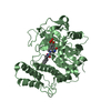
- PDB-5t7x: Crystal structure of HHV-4 EBNA1 DNA binding domain (patient-deri... -
+ Open data

- Basic information
| Entry | Database: PDB / ID: 5t7x | ||||||
|---|---|---|---|---|---|---|---|
| Title | Crystal structure of HHV-4 EBNA1 DNA binding domain (patient-derived, nasopharyngeal carcinoma) bound to DNA | ||||||
 Components Components |
| ||||||
 Keywords Keywords | DNA BINDING PROTEIN/DNA / Dimer / DNA / DNA BINDING PROTEIN-DNA complex | ||||||
| Function / homology |  Function and homology information Function and homology informationviral latency / Hydrolases; Acting on ester bonds; Endodeoxyribonucleases producing 5'-phosphomonoesters / regulation of DNA replication / endonuclease activity / symbiont-mediated suppression of host NF-kappaB cascade / DNA-binding transcription factor activity / hydrolase activity / positive regulation of DNA-templated transcription / host cell nucleus / DNA binding / identical protein binding Similarity search - Function | ||||||
| Biological species |  Human herpesvirus 4 (Epstein-Barr virus) Human herpesvirus 4 (Epstein-Barr virus) | ||||||
| Method |  X-RAY DIFFRACTION / MOLECULAR REPLACEMENT / Resolution: 2.35 Å X-RAY DIFFRACTION / MOLECULAR REPLACEMENT / Resolution: 2.35 Å | ||||||
 Authors Authors | Malecka, K.A. / Messick, T.E. / Lieberman, P.M. | ||||||
 Citation Citation | Journal: Oncotarget / Year: 2017 Title: Carcinoma-risk variant of EBNA1 deregulates Epstein-Barr Virus episomal latency. Authors: Dheekollu, J. / Malecka, K. / Wiedmer, A. / Delecluse, H.J. / Chiang, A.K. / Altieri, D.C. / Messick, T.E. / Lieberman, P.M. | ||||||
| History |
|
- Structure visualization
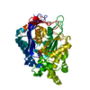
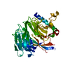
| Structure viewer | Molecule: MolmilJmol/JSmol |
|---|
- Downloads & links
-Download
| PDBx/mmCIF format | 5t7x.cif.gz | 93.5 KB | Display | PDBx/mmCIF format |
|---|---|---|---|---|
| PDB format | pdb5t7x.ent.gz | 66 KB | Display | PDB format |
| PDBx/mmJSON format | 5t7x.json.gz | Tree view | PDBx/mmJSON format | |
| Others |  Other downloads Other downloads |
-Validation report
| Arichive directory | https://data.pdbj.org/pub/pdb/validation_reports/t7/5t7xftp://data.pdbj.org/pub/pdb/validation_reports/t7/5t7x | HTTPS FTP |
|---|
-Related structure data
| Related structure data |  1b3tS S: Starting model for refinement |
|---|---|
| Similar structure data |


-Links
 PDBj
PDBj


- Assembly
| Deposited unit | 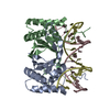
| ||||||||
|---|---|---|---|---|---|---|---|---|---|
| 1 |
| ||||||||
| Unit cell |
|
-Components
| #1: DNA chain | Mass: 5476.554 Da / Num. of mol.: 1 / Source method: obtained synthetically Source: (synth.) Human herpesvirus 4 (strain B95-8) (Epstein-Barr virus (strain B95-8)) | ||
|---|---|---|---|
| #2: DNA chain | Mass: 5556.603 Da / Num. of mol.: 1 / Source method: obtained synthetically Source: (synth.) Human herpesvirus 4 (strain B95-8) (Epstein-Barr virus (strain B95-8)) | ||
| #3: Protein | Mass: 16332.018 Da / Num. of mol.: 2 / Fragment: UNP residues 459-507 Source method: isolated from a genetically manipulated source Details: patient-derived, nasopharyngeal carcinoma Source: (gene. exp.) Human herpesvirus 4 (strain B95-8) (Epstein-Barr virus (strain B95-8))Strain: GD1 / Gene: EBNA1, BKRF1 / Production host:  #4: Water | ChemComp-HOH / |  Mass: 18.015 Da / Num. of mol.: 122 / Source method: isolated from a natural source / Formula: H2O Mass: 18.015 Da / Num. of mol.: 122 / Source method: isolated from a natural source / Formula: H2O |
-Experimental details
-Experiment
| Experiment | Method: X-RAY DIFFRACTION / Number of used crystals: 1 |
|---|
- Sample preparation
| Crystal | Density Matthews: 2.51 Å3/Da / Density % sol: 50.96 % |
|---|---|
| Crystal grow | Temperature: 298 K / Method: vapor diffusion, hanging drop / pH: 7 Details: 0.2 M ammonium acetate, 0.15 M magnesium acetate tetrahydrate, 0.05 M Hepes, pH 7.0, 5% PEG 4000 Temp details: room temperature |
-Data collection
| Diffraction | Mean temperature: 100 K |
|---|---|
| Diffraction source | Source: ROTATING ANODE / Type: RIGAKU / Wavelength: 1.54178 Å |
| Detector | Type: RIGAKU SATURN 944 / Detector: CCD / Date: Aug 27, 2014 |
| Radiation | Protocol: SINGLE WAVELENGTH / Monochromatic (M) / Laue (L): M / Scattering type: x-ray |
| Radiation wavelength | Wavelength: 1.54178 Å / Relative weight: 1 |
| Reflection | Resolution: 2.35→50 Å / Num. obs: 19350 / % possible obs: 98.7 % / Redundancy: 7.2 % / Rmerge(I) obs: 0.198 / Net I/σ(I): 21.9 |
| Reflection shell | Redundancy: 7.2 % / Rmerge(I) obs: 0.879 / Mean I/σ(I) obs: 7.4 / % possible all: 97.6 |
- Processing
| Software |
| |||||||||||||||||||||||||||||||||||||||||||||||||||||||||||||||||||||||||||||||||||||||||||||||||||||||||||||||||||||||
|---|---|---|---|---|---|---|---|---|---|---|---|---|---|---|---|---|---|---|---|---|---|---|---|---|---|---|---|---|---|---|---|---|---|---|---|---|---|---|---|---|---|---|---|---|---|---|---|---|---|---|---|---|---|---|---|---|---|---|---|---|---|---|---|---|---|---|---|---|---|---|---|---|---|---|---|---|---|---|---|---|---|---|---|---|---|---|---|---|---|---|---|---|---|---|---|---|---|---|---|---|---|---|---|---|---|---|---|---|---|---|---|---|---|---|---|---|---|---|---|---|
| Refinement | Method to determine structure: MOLECULAR REPLACEMENT Starting model: 1B3T Resolution: 2.35→23.373 Å / SU ML: 0.29 / Cross valid method: FREE R-VALUE / σ(F): 1.34 / Phase error: 24.79
| |||||||||||||||||||||||||||||||||||||||||||||||||||||||||||||||||||||||||||||||||||||||||||||||||||||||||||||||||||||||
| Solvent computation | Shrinkage radii: 0.9 Å / VDW probe radii: 1.11 Å | |||||||||||||||||||||||||||||||||||||||||||||||||||||||||||||||||||||||||||||||||||||||||||||||||||||||||||||||||||||||
| Refinement step | Cycle: LAST / Resolution: 2.35→23.373 Å
| |||||||||||||||||||||||||||||||||||||||||||||||||||||||||||||||||||||||||||||||||||||||||||||||||||||||||||||||||||||||
| Refine LS restraints |
| |||||||||||||||||||||||||||||||||||||||||||||||||||||||||||||||||||||||||||||||||||||||||||||||||||||||||||||||||||||||
| LS refinement shell |
|
