 Movie
Movie Controller

[English] 日本語
 Yorodumi
- PDB-5bvc: Crystal structure of Lipomyces starkeyi levoglucosan kinase bound... -
+ Open data

- Basic information
| Entry | Database: PDB / ID: 5bvc | ||||||
|---|---|---|---|---|---|---|---|
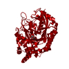
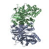
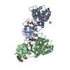
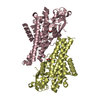
| Title | Crystal structure of Lipomyces starkeyi levoglucosan kinase bound to ADP, magnesium and levoglucosan in an alternate orientation. | ||||||
 Components Components | Levoglucosan kinase | ||||||
 Keywords Keywords | TRANSFERASE / SUGAR KINASE / ATP-BINDING / CARBOHYDRATE METABOLISM / LEVOGLUCOSAN | ||||||
| Function / homology |  Function and homology information Function and homology informationlevoglucosan kinase / amino sugar metabolic process / phosphotransferase activity, alcohol group as acceptor / peptidoglycan turnover / kinase activity / ATP binding / metal ion binding Similarity search - Function | ||||||
| Biological species |  Lipomyces starkeyi (fungus) Lipomyces starkeyi (fungus) | ||||||
| Method |  X-RAY DIFFRACTION / SYNCHROTRON / MOLECULAR REPLACEMENT / Resolution: 2 Å X-RAY DIFFRACTION / SYNCHROTRON / MOLECULAR REPLACEMENT / Resolution: 2 Å | ||||||
 Authors Authors | Bacik, J.P. | ||||||
| Funding support |  United States, 1items United States, 1items
| ||||||
 Citation Citation | Journal: J.Biol.Chem. / Year: 2015 Title: Producing Glucose 6-Phosphate from Cellulosic Biomass: STRUCTURAL INSIGHTS INTO LEVOGLUCOSAN BIOCONVERSION. Authors: Bacik, J.P. / Klesmith, J.R. / Whitehead, T.A. / Jarboe, L.R. / Unkefer, C.J. / Mark, B.L. / Michalczyk, R. | ||||||
| History |
|
- Structure visualization
| Structure viewer | Molecule: MolmilJmol/JSmol |
|---|
- Downloads & links
-Download
| PDBx/mmCIF format | 5bvc.cif.gz | 107.4 KB | Display | PDBx/mmCIF format |
|---|---|---|---|---|
| PDB format | pdb5bvc.ent.gz | 78.9 KB | Display | PDB format |
| PDBx/mmJSON format | 5bvc.json.gz | Tree view | PDBx/mmJSON format | |
| Others |  Other downloads Other downloads |
-Validation report
| Arichive directory | https://data.pdbj.org/pub/pdb/validation_reports/bv/5bvcftp://data.pdbj.org/pub/pdb/validation_reports/bv/5bvc | HTTPS FTP |
|---|
-Related structure data
| Related structure data |  4yh5SC  4zfvC  4zluC  5bsbC S: Starting model for refinement C: citing same article ( |
|---|---|
| Similar structure data |


-Links
 PDBj
PDBj- Assembly
| Deposited unit | 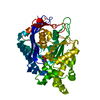
| ||||||||
|---|---|---|---|---|---|---|---|---|---|
| 1 | 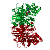
| ||||||||
| Unit cell |
|
-Components
| #1: Protein | Mass: 49395.871 Da / Num. of mol.: 1 Source method: isolated from a genetically manipulated source Source: (gene. exp.) Lipomyces starkeyi (fungus) / Production host:  References: UniProt: B3VI55, Transferases; Transferring phosphorus-containing groups |
|---|---|
| #2: Chemical | ChemComp-ADP /   Mass: 427.201 Da / Num. of mol.: 1 / Source method: obtained synthetically / Formula: C10H15N5O10P2 / Comment: ADP, energy-carrying molecule*YM Mass: 427.201 Da / Num. of mol.: 1 / Source method: obtained synthetically / Formula: C10H15N5O10P2 / Comment: ADP, energy-carrying molecule*YM |
| #3: Chemical | ChemComp-4PW /   Mass: 162.141 Da / Num. of mol.: 1 / Source method: obtained synthetically / Formula: C6H10O5 Mass: 162.141 Da / Num. of mol.: 1 / Source method: obtained synthetically / Formula: C6H10O5 |
| #4: Chemical | ChemComp-MG /   Mass: 24.305 Da / Num. of mol.: 1 / Source method: obtained synthetically / Formula: Mg Mass: 24.305 Da / Num. of mol.: 1 / Source method: obtained synthetically / Formula: Mg |
| #5: Water | ChemComp-HOH /  Mass: 18.015 Da / Num. of mol.: 292 / Source method: isolated from a natural source / Formula: H2O Mass: 18.015 Da / Num. of mol.: 292 / Source method: isolated from a natural source / Formula: H2O |
-Experimental details
-Experiment
| Experiment | Method: X-RAY DIFFRACTION / Number of used crystals: 1 |
|---|
- Sample preparation
| Crystal | Density Matthews: 3.3 Å3/Da / Density % sol: 62.72 % |
|---|---|
| Crystal grow | Temperature: 296 K / Method: vapor diffusion, hanging drop Details: Equal amounts of LGK (25 mg/ml) in crystallization buffer (100 mM NaCl, 0.5 mM TCEP, 20 mM Tris pH 7.5, 2 mM ADP, 4 mM MgCl, 1 mM aluminum nitrate and 10 mM sodium fluoride) and ...Details: Equal amounts of LGK (25 mg/ml) in crystallization buffer (100 mM NaCl, 0.5 mM TCEP, 20 mM Tris pH 7.5, 2 mM ADP, 4 mM MgCl, 1 mM aluminum nitrate and 10 mM sodium fluoride) and crystallization buffer (1.3 M sodium malonate, 0.093 M Bis-Tris Propane pH 7.0) were mixed and and a resultant crystal was soaked with 200 mM levoglucosan. |
-Data collection
| Diffraction | Mean temperature: 100 K |
|---|---|
| Diffraction source | Source: SYNCHROTRON / Site: SSRL / Beamline: BL7-1 / Wavelength: 1.12709 Å |
| Detector | Type: ADSC QUANTUM 315r / Detector: CCD / Date: Jul 8, 2014 |
| Radiation | Protocol: SINGLE WAVELENGTH / Monochromatic (M) / Laue (L): M / Scattering type: x-ray |
| Radiation wavelength | Wavelength: 1.12709 Å / Relative weight: 1 |
| Reflection | Resolution: 2→43.27 Å / Num. obs: 44939 / % possible obs: 98.5 % / Redundancy: 3.3 % / Rmerge(I) obs: 0.1 / Net I/σ(I): 6.2 |
| Reflection shell | Resolution: 2→2.11 Å / Redundancy: 3.4 % / Rmerge(I) obs: 0.592 / Mean I/σ(I) obs: 2 / % possible all: 99.9 |
- Processing
| Software |
| |||||||||||||||||||||||||||||||||||||||||||||||||||||||||||||||||||||||||||||||||||||||||||||||||||||||||||||||||||||||
|---|---|---|---|---|---|---|---|---|---|---|---|---|---|---|---|---|---|---|---|---|---|---|---|---|---|---|---|---|---|---|---|---|---|---|---|---|---|---|---|---|---|---|---|---|---|---|---|---|---|---|---|---|---|---|---|---|---|---|---|---|---|---|---|---|---|---|---|---|---|---|---|---|---|---|---|---|---|---|---|---|---|---|---|---|---|---|---|---|---|---|---|---|---|---|---|---|---|---|---|---|---|---|---|---|---|---|---|---|---|---|---|---|---|---|---|---|---|---|---|---|
| Refinement | Method to determine structure: MOLECULAR REPLACEMENT Starting model: 4YH5 Resolution: 2→43.27 Å / SU ML: 0.22 / Cross valid method: FREE R-VALUE / σ(F): 1.34 / Phase error: 23.92 / Stereochemistry target values: ML
| |||||||||||||||||||||||||||||||||||||||||||||||||||||||||||||||||||||||||||||||||||||||||||||||||||||||||||||||||||||||
| Solvent computation | Shrinkage radii: 0.9 Å / VDW probe radii: 1.11 Å / Solvent model: FLAT BULK SOLVENT MODEL | |||||||||||||||||||||||||||||||||||||||||||||||||||||||||||||||||||||||||||||||||||||||||||||||||||||||||||||||||||||||
| Refinement step | Cycle: LAST / Resolution: 2→43.27 Å
| |||||||||||||||||||||||||||||||||||||||||||||||||||||||||||||||||||||||||||||||||||||||||||||||||||||||||||||||||||||||
| Refine LS restraints |
| |||||||||||||||||||||||||||||||||||||||||||||||||||||||||||||||||||||||||||||||||||||||||||||||||||||||||||||||||||||||
| LS refinement shell |
|
