 Movie
Movie Controller

[English] 日本語
 Yorodumi
- PDB-4zxz: Crystal structure of a highly thermal stable but inactive levoglu... -
+ Open data

- Basic information
| Entry | Database: PDB / ID: 4zxz | |||||||||
|---|---|---|---|---|---|---|---|---|---|---|
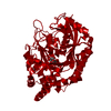
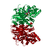
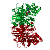
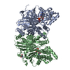
| Title | Crystal structure of a highly thermal stable but inactive levoglucosan kinase. | |||||||||
 Components Components | Levoglucosan kinase | |||||||||
 Keywords Keywords | DE NOVO PROTEIN / Transferase / sugar kinase. | |||||||||
| Function / homology | ATPase, nucleotide binding domain / Nucleotidyltransferase; domain 5 / 2-Layer Sandwich / Alpha Beta Function and homology information Function and homology information | |||||||||
| Biological species |  Lipomyces starkeyi (fungus) Lipomyces starkeyi (fungus) | |||||||||
| Method |  X-RAY DIFFRACTION / MOLECULAR REPLACEMENT / Resolution: 2.2 Å X-RAY DIFFRACTION / MOLECULAR REPLACEMENT / Resolution: 2.2 Å | |||||||||
 Authors Authors | Bacik, J.P. / Klesmith, J.R. / Whitehead, T.A. | |||||||||
| Funding support |  United States, 2items United States, 2items
| |||||||||
 Citation Citation | Journal: Acs Synth Biol / Year: 2015 Title: Comprehensive Sequence-Flux Mapping of a Levoglucosan Utilization Pathway in E. coli. Authors: Klesmith, J.R. / Bacik, J.P. / Michalczyk, R. / Whitehead, T.A. | |||||||||
| History |
|
- Structure visualization
| Structure viewer | Molecule: MolmilJmol/JSmol |
|---|
- Downloads & links
-Download
| PDBx/mmCIF format | 4zxz.cif.gz | 182.1 KB | Display | PDBx/mmCIF format |
|---|---|---|---|---|
| PDB format | pdb4zxz.ent.gz | 143.7 KB | Display | PDB format |
| PDBx/mmJSON format | 4zxz.json.gz | Tree view | PDBx/mmJSON format | |
| Others |  Other downloads Other downloads |
-Validation report
| Arichive directory | https://data.pdbj.org/pub/pdb/validation_reports/zx/4zxzftp://data.pdbj.org/pub/pdb/validation_reports/zx/4zxz | HTTPS FTP |
|---|
-Related structure data
| Related structure data |  4yh5S S: Starting model for refinement |
|---|---|
| Similar structure data |


-Links
 PDBj
PDBj
- Assembly
| Deposited unit | 
| ||||||||
|---|---|---|---|---|---|---|---|---|---|
| 1 |
| ||||||||
| Unit cell |
|
-Components
| #1: Protein | Mass: 49458.941 Da / Num. of mol.: 2 Source method: isolated from a genetically manipulated source Source: (gene. exp.) Lipomyces starkeyi (fungus) / Production host:  #2: Water | ChemComp-HOH / |  Mass: 18.015 Da / Num. of mol.: 394 / Source method: isolated from a natural source / Formula: H2O Mass: 18.015 Da / Num. of mol.: 394 / Source method: isolated from a natural source / Formula: H2O |
|---|
-Experimental details
-Experiment
| Experiment | Method: X-RAY DIFFRACTION |
|---|
- Sample preparation
| Crystal | Density Matthews: 2.67 Å3/Da / Density % sol: 53.94 % |
|---|---|
| Crystal grow | Temperature: 296 K / Method: vapor diffusion, hanging drop / pH: 7.5 Details: Crystals were grown by mixing equal volumes of reservoir buffer containing 18% PEG3350 and 500 mM ammonium tartrate and the LGK construct at 21.5 mg/ml in crystallization buffer containing ...Details: Crystals were grown by mixing equal volumes of reservoir buffer containing 18% PEG3350 and 500 mM ammonium tartrate and the LGK construct at 21.5 mg/ml in crystallization buffer containing 20 mM Tris pH 7.5, 50 mM NaCl and 0.5 mM TCEP. |
-Data collection
| Diffraction | Mean temperature: 296 K |
|---|---|
| Diffraction source | Source: ROTATING ANODE / Type: RIGAKU / Wavelength: 1.5418 Å |
| Detector | Type: RIGAKU / Detector: IMAGE PLATE / Date: Oct 30, 2014 |
| Radiation | Protocol: SINGLE WAVELENGTH / Monochromatic (M) / Laue (L): M / Scattering type: x-ray |
| Radiation wavelength | Wavelength: 1.5418 Å / Relative weight: 1 |
| Reflection | Resolution: 2.2→46.13 Å / Num. obs: 49339 / % possible obs: 90.9 % / Redundancy: 3.6 % / Rmerge(I) obs: 0.147 / Net I/σ(I): 6.9 |
| Reflection shell | Resolution: 2.2→2.32 Å / Redundancy: 2.9 % / Rmerge(I) obs: 0.765 / Mean I/σ(I) obs: 1.6 / % possible all: 79 |
- Processing
| Software |
| |||||||||||||||||||||||||||||||||||||||||||||||||||||||||||||||||||||||||||||||||||||||||||||||||||||||||||||||||||||||||||||||||||||
|---|---|---|---|---|---|---|---|---|---|---|---|---|---|---|---|---|---|---|---|---|---|---|---|---|---|---|---|---|---|---|---|---|---|---|---|---|---|---|---|---|---|---|---|---|---|---|---|---|---|---|---|---|---|---|---|---|---|---|---|---|---|---|---|---|---|---|---|---|---|---|---|---|---|---|---|---|---|---|---|---|---|---|---|---|---|---|---|---|---|---|---|---|---|---|---|---|---|---|---|---|---|---|---|---|---|---|---|---|---|---|---|---|---|---|---|---|---|---|---|---|---|---|---|---|---|---|---|---|---|---|---|---|---|---|
| Refinement | Method to determine structure: MOLECULAR REPLACEMENT Starting model: 4YH5 Resolution: 2.2→46.13 Å / SU ML: 0.25 / Cross valid method: FREE R-VALUE / σ(F): 1.34 / Phase error: 22.13 / Stereochemistry target values: ML
| |||||||||||||||||||||||||||||||||||||||||||||||||||||||||||||||||||||||||||||||||||||||||||||||||||||||||||||||||||||||||||||||||||||
| Solvent computation | Shrinkage radii: 0.9 Å / VDW probe radii: 1.11 Å / Solvent model: FLAT BULK SOLVENT MODEL | |||||||||||||||||||||||||||||||||||||||||||||||||||||||||||||||||||||||||||||||||||||||||||||||||||||||||||||||||||||||||||||||||||||
| Refinement step | Cycle: LAST / Resolution: 2.2→46.13 Å
| |||||||||||||||||||||||||||||||||||||||||||||||||||||||||||||||||||||||||||||||||||||||||||||||||||||||||||||||||||||||||||||||||||||
| Refine LS restraints |
| |||||||||||||||||||||||||||||||||||||||||||||||||||||||||||||||||||||||||||||||||||||||||||||||||||||||||||||||||||||||||||||||||||||
| LS refinement shell |
|
