 Movie
Movie Controller

[English] 日本語
 Yorodumi
- PDB-5t7d: Crystal structure of Streptomyces hygroscopicus bialaphos resista... -
+ Open data

- Basic information
| Entry | Database: PDB / ID: 5t7d | |||||||||||||||||||||
|---|---|---|---|---|---|---|---|---|---|---|---|---|---|---|---|---|---|---|---|---|---|---|
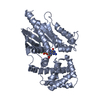
| Title | Crystal structure of Streptomyces hygroscopicus bialaphos resistance (BAR) protein in complex with acetyl coenzyme A | |||||||||||||||||||||
 Components Components | Phosphinothricin N-acetyltransferase | |||||||||||||||||||||
 Keywords Keywords | TRANSFERASE / Gcn5-related N-acetyltransferases Phosphinothricin-deactivating activity | |||||||||||||||||||||
| Function / homology |  Function and homology information Function and homology informationphosphinothricin acetyltransferase / phosphinothricin N-acetyltransferase activity / response to herbicide / response to antibiotic Similarity search - Function | |||||||||||||||||||||
| Biological species |  Streptomyces hygroscopicus (bacteria) Streptomyces hygroscopicus (bacteria) | |||||||||||||||||||||
| Method |  X-RAY DIFFRACTION / SYNCHROTRON / MOLECULAR REPLACEMENT / Resolution: 1.4 Å X-RAY DIFFRACTION / SYNCHROTRON / MOLECULAR REPLACEMENT / Resolution: 1.4 Å | |||||||||||||||||||||
 Authors Authors | Christ, B. / Weng, J.K. | |||||||||||||||||||||
| Funding support |  United States, United States,  Switzerland, United Arab Emirates, 6items Switzerland, United Arab Emirates, 6items
| |||||||||||||||||||||
 Citation Citation | Journal: Nat Plants / Year: 2017 Title: Non-specific activities of the major herbicide-resistance gene BAR. Authors: Christ, B. / Hochstrasser, R. / Guyer, L. / Francisco, R. / Aubry, S. / Hortensteiner, S. / Weng, J.K. | |||||||||||||||||||||
| History |
|
- Structure visualization
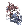
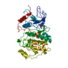
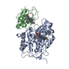
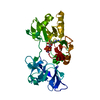
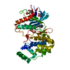
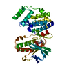
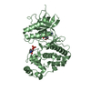
| Structure viewer | Molecule: MolmilJmol/JSmol |
|---|
- Downloads & links
-Download
| PDBx/mmCIF format | 5t7d.cif.gz | 459.9 KB | Display | PDBx/mmCIF format |
|---|---|---|---|---|
| PDB format | pdb5t7d.ent.gz | 387 KB | Display | PDB format |
| PDBx/mmJSON format | 5t7d.json.gz | Tree view | PDBx/mmJSON format | |
| Others |  Other downloads Other downloads |
-Validation report
| Summary document | 5t7d_validation.pdf.gz | 467.8 KB | Display | wwPDB validaton report |
|---|---|---|---|---|
| Full document | 5t7d_full_validation.pdf.gz | 475 KB | Display | |
| Data in XML | 5t7d_validation.xml.gz | 3.2 KB | Display | |
| Data in CIF | 5t7d_validation.cif.gz | 16.5 KB | Display | |
| Arichive directory | https://data.pdbj.org/pub/pdb/validation_reports/t7/5t7dftp://data.pdbj.org/pub/pdb/validation_reports/t7/5t7d | HTTPS FTP |
-Related structure data


-Links
 PDBj
PDBj

- Assembly
| Deposited unit | 
| ||||||||
|---|---|---|---|---|---|---|---|---|---|
| 1 | 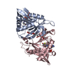
| ||||||||
| 2 | 
| ||||||||
| Unit cell |
|
-Components
| #1: Protein | Mass: 21281.051 Da / Num. of mol.: 4 / Fragment: UNP residues 7-181 Source method: isolated from a genetically manipulated source Source: (gene. exp.) Streptomyces hygroscopicus (bacteria) / Gene: bar / Plasmid: pProExHta / Production host: References: UniProt: P16426, phosphinothricin acetyltransferase #2: Chemical | ChemComp-ACO /   Mass: 809.571 Da / Num. of mol.: 4 Mass: 809.571 Da / Num. of mol.: 4Source method: isolated from a genetically manipulated source Formula: C23H38N7O17P3S #3: Chemical | ChemComp-ACT /   Mass: 59.044 Da / Num. of mol.: 8 Mass: 59.044 Da / Num. of mol.: 8Source method: isolated from a genetically manipulated source Formula: C2H3O2 #4: Water | ChemComp-HOH / |  Mass: 18.015 Da / Num. of mol.: 1073 / Source method: isolated from a natural source / Formula: H2O Mass: 18.015 Da / Num. of mol.: 1073 / Source method: isolated from a natural source / Formula: H2O |
|---|
-Experimental details
-Experiment
| Experiment | Method: X-RAY DIFFRACTION / Number of used crystals: 1 |
|---|
- Sample preparation
| Crystal | Density Matthews: 2.32 Å3/Da / Density % sol: 47.08 % |
|---|---|
| Crystal grow | Temperature: 293 K / Method: vapor diffusion, hanging drop Details: BAR protein was incubated with 1 mM acetyl-CoA for >2 hour prior to setting crystal trays. Crystals of BAR were obtained after 3 days at 20C in hanging drops containing 1 uL of protein ...Details: BAR protein was incubated with 1 mM acetyl-CoA for >2 hour prior to setting crystal trays. Crystals of BAR were obtained after 3 days at 20C in hanging drops containing 1 uL of protein solution (7.5 mg/mL) and 1 uL of reservoir solution (0.18 M calcium acetate, 0.1 M Tris-HCl pH 7, 18% (w/v) PEG 3000, 0.2% (v/v) N-nonyl Beta-D-glucopyranoside, 1 mM acetyl-CoA). Crystals were frozen in reservoir solution supplemented with 15% (v/v) ethylene glycol. |
-Data collection
| Diffraction | Mean temperature: 90 K |
|---|---|
| Diffraction source | Source: SYNCHROTRON / Site: APS / Beamline: 24-ID-C / Wavelength: 0.987 Å |
| Detector | Type: DECTRIS PILATUS 6M-F / Detector: PIXEL / Date: Aug 8, 2015 |
| Radiation | Protocol: SINGLE WAVELENGTH / Monochromatic (M) / Laue (L): M / Scattering type: x-ray |
| Radiation wavelength | Wavelength: 0.987 Å / Relative weight: 1 |
| Reflection | Resolution: 1.4→44.79 Å / Num. obs: 238978 / % possible obs: 89.48 % / Redundancy: 1.8 % / Biso Wilson estimate: 14.16 Å2 / CC1/2: 0.998 / Net I/σ(I): 8.84 |
- Processing
| Software |
| |||||||||||||||||||||||||||||||||||||||||||||||||||||||||||||||||||||||||||||||||||||||||||||||||||||||||||||||||||||||||||||||||||||||||||||||||||||||||||||||||||||||||||||||||||||||||||||
|---|---|---|---|---|---|---|---|---|---|---|---|---|---|---|---|---|---|---|---|---|---|---|---|---|---|---|---|---|---|---|---|---|---|---|---|---|---|---|---|---|---|---|---|---|---|---|---|---|---|---|---|---|---|---|---|---|---|---|---|---|---|---|---|---|---|---|---|---|---|---|---|---|---|---|---|---|---|---|---|---|---|---|---|---|---|---|---|---|---|---|---|---|---|---|---|---|---|---|---|---|---|---|---|---|---|---|---|---|---|---|---|---|---|---|---|---|---|---|---|---|---|---|---|---|---|---|---|---|---|---|---|---|---|---|---|---|---|---|---|---|---|---|---|---|---|---|---|---|---|---|---|---|---|---|---|---|---|---|---|---|---|---|---|---|---|---|---|---|---|---|---|---|---|---|---|---|---|---|---|---|---|---|---|---|---|---|---|---|---|---|
| Refinement | Method to determine structure: MOLECULAR REPLACEMENT / Resolution: 1.4→44.788 Å / SU ML: 0.15 / Cross valid method: FREE R-VALUE / σ(F): 1.69 / Phase error: 21.52 / Stereochemistry target values: ML
| |||||||||||||||||||||||||||||||||||||||||||||||||||||||||||||||||||||||||||||||||||||||||||||||||||||||||||||||||||||||||||||||||||||||||||||||||||||||||||||||||||||||||||||||||||||||||||||
| Solvent computation | Shrinkage radii: 0.9 Å / VDW probe radii: 1.11 Å / Solvent model: FLAT BULK SOLVENT MODEL | |||||||||||||||||||||||||||||||||||||||||||||||||||||||||||||||||||||||||||||||||||||||||||||||||||||||||||||||||||||||||||||||||||||||||||||||||||||||||||||||||||||||||||||||||||||||||||||
| Refinement step | Cycle: LAST / Resolution: 1.4→44.788 Å
| |||||||||||||||||||||||||||||||||||||||||||||||||||||||||||||||||||||||||||||||||||||||||||||||||||||||||||||||||||||||||||||||||||||||||||||||||||||||||||||||||||||||||||||||||||||||||||||
| Refine LS restraints |
| |||||||||||||||||||||||||||||||||||||||||||||||||||||||||||||||||||||||||||||||||||||||||||||||||||||||||||||||||||||||||||||||||||||||||||||||||||||||||||||||||||||||||||||||||||||||||||||
| LS refinement shell |
|
