 Movie
Movie Controller

[English] 日本語
 Yorodumi
- PDB-5t16: Crystal structure of yeast RNase III (Rnt1p) complexed with a non... -
+ Open data

- Basic information
| Entry | Database: PDB / ID: 5t16 | ||||||
|---|---|---|---|---|---|---|---|
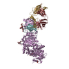
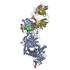
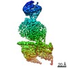
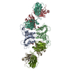
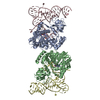
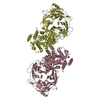
| Title | Crystal structure of yeast RNase III (Rnt1p) complexed with a non-hydrolyzable RNA substrate analog | ||||||
 Components Components |
| ||||||
 Keywords Keywords | hydrolase/rna / Rnt1p / RNase III / substrate-loaded complex / hydrolase-rna complex | ||||||
| Function / homology |  Function and homology information Function and homology informationbox C/D sno(s)RNA processing / box H/ACA sno(s)RNA processing / regulation of fungal-type cell wall organization / termination of RNA polymerase II transcription, exosome-dependent / U1 snRNA 3'-end processing / U5 snRNA 3'-end processing / U4 snRNA 3'-end processing / ribonuclease III / ribonuclease III activity / termination of RNA polymerase II transcription ...box C/D sno(s)RNA processing / box H/ACA sno(s)RNA processing / regulation of fungal-type cell wall organization / termination of RNA polymerase II transcription, exosome-dependent / U1 snRNA 3'-end processing / U5 snRNA 3'-end processing / U4 snRNA 3'-end processing / ribonuclease III / ribonuclease III activity / termination of RNA polymerase II transcription / rRNA transcription / rRNA processing / double-stranded RNA binding / chromatin organization / nucleolus / nucleoplasm / nucleus Similarity search - Function | ||||||
| Biological species |  | ||||||
| Method |  X-RAY DIFFRACTION / SYNCHROTRON / MOLECULAR REPLACEMENT / Resolution: 2.783 Å X-RAY DIFFRACTION / SYNCHROTRON / MOLECULAR REPLACEMENT / Resolution: 2.783 Å | ||||||
 Authors Authors | Song, H. / Ji, X. | ||||||
 Citation Citation | Journal: Structure / Year: 2017 Title: The Functional Cycle of Rnt1p: Five Consecutive Steps of Double-Stranded RNA Processing by a Eukaryotic RNase III. Authors: Song, H. / Fang, X. / Jin, L. / Shaw, G.X. / Wang, Y.X. / Ji, X. #1: Journal: Mol. Cell / Year: 2014Title: Structure of a eukaryotic RNase III postcleavage complex reveals a double-ruler mechanism for substrate selection. Authors: Liang, Y.H. / Lavoie, M. / Comeau, M.A. / Abou Elela, S. / Ji, X. | ||||||
| History |
|
- Structure visualization
| Structure viewer | Molecule: MolmilJmol/JSmol |
|---|
- Downloads & links
-Download
| PDBx/mmCIF format | 5t16.cif.gz | 491.2 KB | Display | PDBx/mmCIF format |
|---|---|---|---|---|
| PDB format | pdb5t16.ent.gz | 394.3 KB | Display | PDB format |
| PDBx/mmJSON format | 5t16.json.gz | Tree view | PDBx/mmJSON format | |
| Others |  Other downloads Other downloads |
-Validation report
| Arichive directory | https://data.pdbj.org/pub/pdb/validation_reports/t1/5t16ftp://data.pdbj.org/pub/pdb/validation_reports/t1/5t16 | HTTPS FTP |
|---|
-Related structure data
| Related structure data | 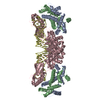 4oogS S: Starting model for refinement |
|---|---|
| Similar structure data |


-Links
 PDBj
PDBj


- Assembly
| Deposited unit | 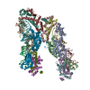
| ||||||||
|---|---|---|---|---|---|---|---|---|---|
| 1 | 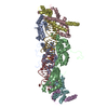
| ||||||||
| 2 | 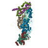
| ||||||||
| Unit cell |
|
-Components
| #1: Protein | Mass: 31481.312 Da / Num. of mol.: 4 / Fragment: unp residues 184-499 Source method: isolated from a genetically manipulated source Source: (gene. exp.) Strain: ATCC 204508 / S288c / Gene: RNT1, YMR239C, YM9408.01C, YM9959.21 / Production host:  #2: Protein | Mass: 13975.908 Da / Num. of mol.: 8 / Fragment: unp residues 41-199 Source method: isolated from a genetically manipulated source Source: (gene. exp.) Strain: ATCC 204508 / S288c / Gene: RNT1, YMR239C, YM9408.01C, YM9959.21 / Production host: #3: RNA chain | Mass: 10940.617 Da / Num. of mol.: 4 / Source method: obtained synthetically #4: Chemical | ChemComp-EDO /   Mass: 62.068 Da / Num. of mol.: 10 / Source method: obtained synthetically / Formula: C2H6O2 Mass: 62.068 Da / Num. of mol.: 10 / Source method: obtained synthetically / Formula: C2H6O2#5: Water | ChemComp-HOH / |  Mass: 18.015 Da / Num. of mol.: 358 / Source method: isolated from a natural source / Formula: H2O Mass: 18.015 Da / Num. of mol.: 358 / Source method: isolated from a natural source / Formula: H2O |
|---|
-Experimental details
-Experiment
| Experiment | Method: X-RAY DIFFRACTION / Number of used crystals: 1 |
|---|
- Sample preparation
| Crystal | Density Matthews: 3.19 Å3/Da / Density % sol: 61.49 % |
|---|---|
| Crystal grow | Temperature: 293 K / Method: vapor diffusion, sitting drop / Details: 20% PEG 3350, 0.2 M di ammonium citrate |
-Data collection
| Diffraction | Mean temperature: 100 K |
|---|---|
| Diffraction source | Source: SYNCHROTRON / Site: APS  / Beamline: 22-ID / Wavelength: 1 Å / Beamline: 22-ID / Wavelength: 1 Å |
| Detector | Type: RAYONIX MX300HE / Detector: CCD / Date: Nov 6, 2015 |
| Radiation | Monochromator: double crystal / Protocol: SINGLE WAVELENGTH / Monochromatic (M) / Laue (L): M / Scattering type: x-ray |
| Radiation wavelength | Wavelength: 1 Å / Relative weight: 1 |
| Reflection | Resolution: 2.78→38.99 Å / Num. obs: 86174 / % possible obs: 99.3 % / Redundancy: 4.5 % / Net I/σ(I): 6.79 |
| Reflection shell | Resolution: 2.78→2.9 Å / Redundancy: 3.8 % / % possible all: 96 |
- Processing
| Software |
| ||||||||||||||||||||||||||||||||||||||||||||||||||||||||
|---|---|---|---|---|---|---|---|---|---|---|---|---|---|---|---|---|---|---|---|---|---|---|---|---|---|---|---|---|---|---|---|---|---|---|---|---|---|---|---|---|---|---|---|---|---|---|---|---|---|---|---|---|---|---|---|---|---|
| Refinement | Method to determine structure: MOLECULAR REPLACEMENT Starting model: 4OOG Resolution: 2.783→38.985 Å / SU ML: 0.44 / Cross valid method: FREE R-VALUE / σ(F): 1.34 / Phase error: 35.92
| ||||||||||||||||||||||||||||||||||||||||||||||||||||||||
| Solvent computation | Shrinkage radii: 0.9 Å / VDW probe radii: 1.11 Å | ||||||||||||||||||||||||||||||||||||||||||||||||||||||||
| Refinement step | Cycle: LAST / Resolution: 2.783→38.985 Å
| ||||||||||||||||||||||||||||||||||||||||||||||||||||||||
| LS refinement shell |
|
