 Movie
Movie Controller

[English] 日本語
 Yorodumi
- PDB-5suo: Crystal structure of N-glycan transport solute binding protein (N... -
+ Open data

- Basic information
| Entry | Database: PDB / ID: 5suo | ||||||
|---|---|---|---|---|---|---|---|
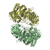
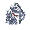
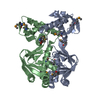
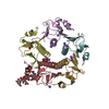
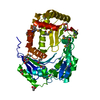
| Title | Crystal structure of N-glycan transport solute binding protein (NgtS) from Streptococcus pneumoniae | ||||||
 Components Components | Extracellular solute-binding protein | ||||||
 Keywords Keywords | PROTEIN BINDING / Solute binding protein / alpha/beta domain | ||||||
| Function / homology | IODIDE ION / :  Function and homology information Function and homology information | ||||||
| Biological species |   Streptococcus pneumoniae (bacteria) Streptococcus pneumoniae (bacteria) | ||||||
| Method |  X-RAY DIFFRACTION / SAD / Resolution: 2.1 Å X-RAY DIFFRACTION / SAD / Resolution: 2.1 Å | ||||||
 Authors Authors | Robb, M. / Boraston, A.B. | ||||||
| Funding support |  Canada, 1items Canada, 1items
| ||||||
 Citation Citation | Journal: PLoS Pathog. / Year: 2017 Title: Molecular Characterization of N-glycan Degradation and Transport in Streptococcus pneumoniae and Its Contribution to Virulence. Authors: Robb, M. / Hobbs, J.K. / Woodiga, S.A. / Shapiro-Ward, S. / Suits, M.D. / McGregor, N. / Brumer, H. / Yesilkaya, H. / King, S.J. / Boraston, A.B. | ||||||
| History |
|
- Structure visualization
| Structure viewer | Molecule: MolmilJmol/JSmol |
|---|
- Downloads & links
-Download
| PDBx/mmCIF format | 5suo.cif.gz | 106.6 KB | Display | PDBx/mmCIF format |
|---|---|---|---|---|
| PDB format | pdb5suo.ent.gz | 79.1 KB | Display | PDB format |
| PDBx/mmJSON format | 5suo.json.gz | Tree view | PDBx/mmJSON format | |
| Others |  Other downloads Other downloads |
-Validation report
| Arichive directory | https://data.pdbj.org/pub/pdb/validation_reports/su/5suoftp://data.pdbj.org/pub/pdb/validation_reports/su/5suo | HTTPS FTP |
|---|
-Related structure data


-Links
 PDBj
PDBj- Assembly
| Deposited unit | 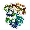
| ||||||||
|---|---|---|---|---|---|---|---|---|---|
| 1 |
| ||||||||
| Unit cell |
|
-Components
| #1: Protein | Mass: 50671.984 Da / Num. of mol.: 1 Source method: isolated from a genetically manipulated source Source: (gene. exp.) Streptococcus pneumoniae (bacteria) / Gene: ERS020541_01426, ERS558328_01721 / Plasmid: pET28a / Production host: | ||
|---|---|---|---|
| #2: Chemical | ChemComp-IOD /   Mass: 126.904 Da / Num. of mol.: 21 / Source method: obtained synthetically / Formula: I Mass: 126.904 Da / Num. of mol.: 21 / Source method: obtained synthetically / Formula: I#3: Water | ChemComp-HOH / |  Mass: 18.015 Da / Num. of mol.: 227 / Source method: isolated from a natural source / Formula: H2O Mass: 18.015 Da / Num. of mol.: 227 / Source method: isolated from a natural source / Formula: H2O |
-Experimental details
-Experiment
| Experiment | Method: X-RAY DIFFRACTION / Number of used crystals: 1 |
|---|
- Sample preparation
| Crystal | Density Matthews: 2.24 Å3/Da / Density % sol: 45.14 % |
|---|---|
| Crystal grow | Temperature: 291 K / Method: vapor diffusion, hanging drop / pH: 8.5 Details: 29 % PEG 1500, 11 % 2-methyl-2,4-pentanediol, 0.1 M Tris |
-Data collection
| Diffraction | Mean temperature: 100 K |
|---|---|
| Diffraction source | Source: ROTATING ANODE / Type: RIGAKU MICROMAX-002 / Wavelength: 1.54187 Å |
| Detector | Type: RIGAKU RAXIS IV++ / Detector: IMAGE PLATE / Date: Mar 20, 2013 |
| Radiation | Protocol: SINGLE WAVELENGTH / Monochromatic (M) / Laue (L): M / Scattering type: x-ray |
| Radiation wavelength | Wavelength: 1.54187 Å / Relative weight: 1 |
| Reflection | Resolution: 1.73→92.79 Å / Num. obs: 25262 / % possible obs: 99.9 % / Redundancy: 4.7 % / Net I/σ(I): 25.4 |
-Phasing
| Phasing | Method: SAD |
|---|
- Processing
| Software |
| ||||||||||||||||||||||||||||||||||||||||||||||||||||||||||||
|---|---|---|---|---|---|---|---|---|---|---|---|---|---|---|---|---|---|---|---|---|---|---|---|---|---|---|---|---|---|---|---|---|---|---|---|---|---|---|---|---|---|---|---|---|---|---|---|---|---|---|---|---|---|---|---|---|---|---|---|---|---|
| Refinement | Method to determine structure: SAD / Resolution: 2.1→92.79 Å / Cor.coef. Fo:Fc: 0.923 / Cor.coef. Fo:Fc free: 0.885 / SU B: 5.504 / SU ML: 0.148 / Cross valid method: THROUGHOUT / σ(F): 0 / ESU R: 0.272 / ESU R Free: 0.215 / Details: U VALUES : REFINED INDIVIDUALLY
| ||||||||||||||||||||||||||||||||||||||||||||||||||||||||||||
| Solvent computation | Ion probe radii: 0.8 Å / Shrinkage radii: 0.8 Å / VDW probe radii: 1.2 Å | ||||||||||||||||||||||||||||||||||||||||||||||||||||||||||||
| Displacement parameters | Biso max: 87.09 Å2 / Biso mean: 19.711 Å2 / Biso min: 4.59 Å2
| ||||||||||||||||||||||||||||||||||||||||||||||||||||||||||||
| Refinement step | Cycle: final / Resolution: 2.1→92.79 Å
| ||||||||||||||||||||||||||||||||||||||||||||||||||||||||||||
| Refine LS restraints |
| ||||||||||||||||||||||||||||||||||||||||||||||||||||||||||||
| LS refinement shell | Resolution: 2.1→2.155 Å / Total num. of bins used: 20
|
