 Movie
Movie Controller

[English] 日本語
 Yorodumi
- PDB-5sga: Structures of product and inhibitor complexes of Streptomyces gri... -
+ Open data

- Basic information
| Entry | Database: PDB / ID: 5sga | ||||||
|---|---|---|---|---|---|---|---|
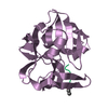
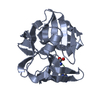
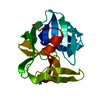
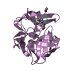
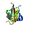
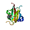
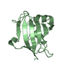
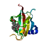
| Title | Structures of product and inhibitor complexes of Streptomyces griseus protease a at 1.8 Angstroms resolution. a model for serine protease catalysis | ||||||
 Components Components |
| ||||||
 Keywords Keywords | HYDROLASE/HYDROLASE INHIBITOR / SERINE PROTEINASE / HYDROLASE-HYDROLASE INHIBITOR complex | ||||||
| Function / homology |  Function and homology information Function and homology informationstreptogrisin A / serine-type endopeptidase activity / proteolysis / extracellular region Similarity search - Function | ||||||
| Biological species |  Streptomyces griseus (bacteria) Streptomyces griseus (bacteria) | ||||||
| Method |  X-RAY DIFFRACTION / Resolution: 1.8 Å X-RAY DIFFRACTION / Resolution: 1.8 Å | ||||||
 Authors Authors | Sielecki, A.R. / James, M.N.G. | ||||||
 Citation Citation | Journal: J.Mol.Biol. / Year: 1980 Title: Structures of product and inhibitor complexes of Streptomyces griseus protease A at 1.8 A resolution. A model for serine protease catalysis. Authors: James, M.N. / Sielecki, A.R. / Brayer, G.D. / Delbaere, L.T. / Bauer, C.A. #1: Journal: Proceedings of the Daresbury Study Weekend: Refinement of Protein StructuresYear: 1981 Title: The Importance of Refined Structures to the Understanding of Enzyme Action Authors: Sielecki, A.R. / James, M.N.G. #2: Journal: J.Mol.Biol. / Year: 1979Title: Protein Structure Refinement. Streptomyces Griseus Serine Protease A at 1.8 Angstroms Resolution Authors: Sielecki, A.R. / Hendrickson, W.A. / Broughton, C.G. / Delbaere, L.T.J. / Brayer, G.D. / James, M.N.G. #3: Journal: J.Mol.Biol. / Year: 1985Title: Electron Density Calculations as an Extension of Protein Structure Refinement. Streptomyces Griseus Protease at 1.5 Angstroms Resolution Authors: Moult, J. / Sussman, F. / James, M.N.G. #4: Journal: Proc.Natl.Acad.Sci.USA / Year: 1979Title: Crystallographic and Kinetic Investigations of the Covalent Complex Formed by a Specific Tetrapeptide Aldehyde and the Serine Protease from Streptomyces Griseus Authors: Brayer, G.D. / Delbaere, L.T.J. / James, M.N.G. / Bauer, C.-A. / Thompson, R.C. #5: Journal: J.Mol.Biol. / Year: 1978Title: Molecular Structure of Crystalline Streptomyces Griseus Protease A at 2.8 Angstroms Resolution. II. Molecular Conformation, Comparison with Alpha-Chymotrypsin and Active-Site Geometry Authors: Brayer, G.D. / Delbaere, L.T.J. / James, M.N.G. #6: Journal: J.Mol.Biol. / Year: 1978Title: Molecular Structure of Crystalline Streptomyces Griseus Protease A at 2.8 Angstroms Resolution. I. Crystallization, Data Collection and Structural Analysis Authors: Brayer, G.D. / Delbaere, L.T.J. / James, M.N.G. #7: Journal: Can.J.Biochem. / Year: 1978Title: Amino Acid Sequence Alignment of Bacterial and Mammalian Pancreatic Serine Proteases Based on Topological Equivalences Authors: James, M.N.G. / Delbaere, L.T.J. / Brayer, G.D. #8: Journal: Nature / Year: 1975Title: Tertiary Structural Differences between Microbial Serine Proteases and Pancreatic Serine Enzymes Authors: Delbaere, L.T.J. / Hutcheon, W.L.B. / James, M.N.G. / Thiessen, W.E. #9: Journal: J.Mol.Biol. / Year: 1980Title: Structure of the Complex Formed between the Bacterial-Produced Inhibitor Chymostatin and the Serine Enzyme Streptomyces Griseus Protease A Authors: Delbaere, L.T.J. / Brayer, G.D. #10: Journal: J.Mol.Biol. / Year: 1985Title: Refined Structure of Alpha-Lytic Protease at 1.7 Angstroms Resolution: Analysis of Hydrogen Bonding and Solvent Structure Authors: Fujinaga, M. / Delbaere, L.T.J. / Brayer, G.D. / James, M.N.G. | ||||||
| History |
|
- Structure visualization
| Structure viewer | Molecule: MolmilJmol/JSmol |
|---|
- Downloads & links
-Download
| PDBx/mmCIF format | 5sga.cif.gz | 50.8 KB | Display | PDBx/mmCIF format |
|---|---|---|---|---|
| PDB format | pdb5sga.ent.gz | 35.1 KB | Display | PDB format |
| PDBx/mmJSON format | 5sga.json.gz | Tree view | PDBx/mmJSON format | |
| Others |  Other downloads Other downloads |
-Validation report
| Arichive directory | https://data.pdbj.org/pub/pdb/validation_reports/sg/5sgaftp://data.pdbj.org/pub/pdb/validation_reports/sg/5sga | HTTPS FTP |
|---|
-Related structure data


-Links
 PDBj
PDBj- Assembly
| Deposited unit | 
| |||||||||||||||
|---|---|---|---|---|---|---|---|---|---|---|---|---|---|---|---|---|
| 1 |
| |||||||||||||||
| Unit cell |
| |||||||||||||||
| Atom site foot note | 1: THE SIDE CHAIN OF ARG E 221 IS VERY DISORDERED. COORDINATES FOR THE ATOMS BEYOND CB HAVE BEEN OMITTED. 2: RESIDUE E 99A IS A CIS-PROLINE. 3: SOLVENT 229, ALTHOUGH REFINED AS AN OXYGEN ATOM, HAS BEEN INTERPRETED AS A NA+ ION. SEE REFERENCE 2 ABOVE FOR FURTHER DETAILS. | |||||||||||||||
| Components on special symmetry positions |
|
-Components
| #1: Protein | Mass: 18016.625 Da / Num. of mol.: 1 Source method: isolated from a genetically manipulated source Source: (gene. exp.) Streptomyces griseus (bacteria) / References: UniProt: P00776 |
|---|---|
| #2: Protein/peptide | Mass: 472.534 Da / Num. of mol.: 1 Source method: isolated from a genetically manipulated source |
| #3: Water | ChemComp-HOH /  Mass: 18.015 Da / Num. of mol.: 185 / Source method: isolated from a natural source / Formula: H2O Mass: 18.015 Da / Num. of mol.: 185 / Source method: isolated from a natural source / Formula: H2O |
| Has protein modification | Y |
-Experimental details
-Experiment
| Experiment | Method: X-RAY DIFFRACTION |
|---|
- Sample preparation
| Crystal | Density Matthews: 2.25 Å3/Da / Density % sol: 45.41 % | ||||||||||||||||||
|---|---|---|---|---|---|---|---|---|---|---|---|---|---|---|---|---|---|---|---|
| Crystal grow | *PLUS pH: 4.1 / Method: equilibrium dialysis | ||||||||||||||||||
| Components of the solutions | *PLUS
|
-Data collection
| Radiation | Scattering type: x-ray |
|---|---|
| Radiation wavelength | Relative weight: 1 |
| Reflection | *PLUS Highest resolution: 1.8 Å / Num. obs: 15254 / % possible obs: 80.7 % / Observed criterion σ(I): 3 / Num. measured all: 16136 / Rmerge(I) obs: 0.085 / Biso Wilson estimate: 12.1 Å2 |
- Processing
| Software | Name: PROLSQ / Classification: refinement | ||||||||||||||||||||||||||||||||||||||||||||||||||||||||||||||||||||||||||||||||||||
|---|---|---|---|---|---|---|---|---|---|---|---|---|---|---|---|---|---|---|---|---|---|---|---|---|---|---|---|---|---|---|---|---|---|---|---|---|---|---|---|---|---|---|---|---|---|---|---|---|---|---|---|---|---|---|---|---|---|---|---|---|---|---|---|---|---|---|---|---|---|---|---|---|---|---|---|---|---|---|---|---|---|---|---|---|---|
| Refinement | Resolution: 1.8→10 Å / Rfactor obs: 0.116 | ||||||||||||||||||||||||||||||||||||||||||||||||||||||||||||||||||||||||||||||||||||
| Refinement step | Cycle: LAST / Resolution: 1.8→10 Å
| ||||||||||||||||||||||||||||||||||||||||||||||||||||||||||||||||||||||||||||||||||||
| Refine LS restraints |
| ||||||||||||||||||||||||||||||||||||||||||||||||||||||||||||||||||||||||||||||||||||
| Refinement | *PLUS Highest resolution: 1.8 Å / Lowest resolution: 10 Å / Rfactor obs: 0.116 / Num. reflection obs: 12166 | ||||||||||||||||||||||||||||||||||||||||||||||||||||||||||||||||||||||||||||||||||||
| Solvent computation | *PLUS | ||||||||||||||||||||||||||||||||||||||||||||||||||||||||||||||||||||||||||||||||||||
| Displacement parameters | *PLUS Biso mean: 14.3 Å2 |
