 Movie
Movie Controller

[English] 日本語
 Yorodumi
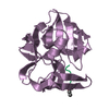
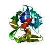
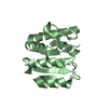
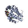
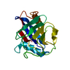
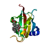
- PDB-1sgc: THE 1.8 ANGSTROMS STRUCTURE OF THE COMPLEX BETWEEN CHYMOSTATIN AN... -
+ Open data

- Basic information
| Entry | Database: PDB / ID: 1sgc | |||||||||
|---|---|---|---|---|---|---|---|---|---|---|
| Title | THE 1.8 ANGSTROMS STRUCTURE OF THE COMPLEX BETWEEN CHYMOSTATIN AND STREPTOMYCES GRISEUS PROTEASE A. A MODEL FOR SERINE PROTEASE CATALYTIC TETRAHEDRAL INTERMEDIATES | |||||||||
 Components Components |
| |||||||||
 Keywords Keywords | HYDROLASE/HYDROLASE INHIBITOR / HYDROLASE (SERINE PROTEINASE) / HYDROLASE-HYDROLASE INHIBITOR COMPLEX | |||||||||
| Function / homology |  Function and homology information Function and homology informationstreptogrisin A / serine-type endopeptidase activity / proteolysis / extracellular region Similarity search - Function | |||||||||
| Biological species |  Streptomyces griseus (bacteria)Streptomyces hygroscopicus (bacteria) Streptomyces griseus (bacteria)Streptomyces hygroscopicus (bacteria)MC521-C8 | |||||||||
| Method |  X-RAY DIFFRACTION / Resolution: 1.8 Å X-RAY DIFFRACTION / Resolution: 1.8 Å | |||||||||
 Authors Authors | Delbaere, L.T.J. / Brayer, G.D. | |||||||||
 Citation Citation | Journal: J.Mol.Biol. / Year: 1985 Title: The 1.8 A structure of the complex between chymostatin and Streptomyces griseus protease A. A model for serine protease catalytic tetrahedral intermediates. Authors: Delbaere, L.T. / Brayer, G.D. #1: Journal: J.Mol.Biol. / Year: 1980Title: Structure of the Complex Formed between the Bacterial-Produced Inhibitor Chymostatin and the Serine Enzyme Streptomyces Griseus Protease A Authors: Delbaere, L.T.J. / Brayer, G.D. | |||||||||
| History |
|
- Structure visualization
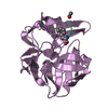
| Structure viewer | Molecule: MolmilJmol/JSmol |
|---|
- Downloads & links
-Download
| PDBx/mmCIF format | 1sgc.cif.gz | 52.3 KB | Display | PDBx/mmCIF format |
|---|---|---|---|---|
| PDB format | pdb1sgc.ent.gz | 35.9 KB | Display | PDB format |
| PDBx/mmJSON format | 1sgc.json.gz | Tree view | PDBx/mmJSON format | |
| Others |  Other downloads Other downloads |
-Validation report
| Arichive directory | https://data.pdbj.org/pub/pdb/validation_reports/sg/1sgcftp://data.pdbj.org/pub/pdb/validation_reports/sg/1sgc | HTTPS FTP |
|---|
-Related structure data
| Similar structure data |
|---|


-Links
 PDBj
PDBj- Assembly
| Deposited unit | 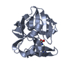
| |||||||||||||||
|---|---|---|---|---|---|---|---|---|---|---|---|---|---|---|---|---|
| 1 |
| |||||||||||||||
| Unit cell |
| |||||||||||||||
| Atom site foot note | 1: RESIDUE 99A IS A CIS-PROLINE. 2: THE SIDE CHAIN ATOMS OF ARG 221 BEYOND CB WERE NOT WELL DEFINED AND, THEREFORE, ARE NOT INCLUDED IN THIS ENTRY. | |||||||||||||||
| Components on special symmetry positions |
|
-Components
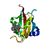
| #1: Protein | Mass: 18016.625 Da / Num. of mol.: 1 Source method: isolated from a genetically manipulated source Source: (gene. exp.) Streptomyces griseus (bacteria) / References: UniProt: P00776 |
|---|---|
| #2: Protein/peptide | 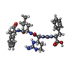  Type: Oligopeptide / Class: Enzyme inhibitor / Mass: 607.701 Da / Num. of mol.: 1 / Source method: obtained synthetically / Source: (synth.) Streptomyces hygroscopicus, MC521-C8 / References: Chymostatin A Type: Oligopeptide / Class: Enzyme inhibitor / Mass: 607.701 Da / Num. of mol.: 1 / Source method: obtained synthetically / Source: (synth.) Streptomyces hygroscopicus, MC521-C8 / References: Chymostatin A |
| #3: Water | ChemComp-HOH /  Mass: 18.015 Da / Num. of mol.: 217 / Source method: isolated from a natural source / Formula: H2O Mass: 18.015 Da / Num. of mol.: 217 / Source method: isolated from a natural source / Formula: H2O |
| Compound details | THE INHIBITOR CHYMOSTATIN A HAS LAST RESIDUE PHENYLALANINAL REPRESENTED WITH ALTERNATE ...THE INHIBITOR CHYMOSTATI |
| Has protein modification | Y |
-Experimental details
-Experiment
| Experiment | Method: X-RAY DIFFRACTION |
|---|
- Sample preparation
| Crystal | Density Matthews: 2.3 Å3/Da / Density % sol: 46.45 % |
|---|---|
| Crystal grow | *PLUS Method: other / Details: Delbaere, L.T.J., (1980) J. Mol. Biol., 139, 45. |
-Data collection
| Radiation | Scattering type: x-ray |
|---|---|
| Radiation wavelength | Relative weight: 1 |
| Reflection | *PLUS Highest resolution: 1.8 Å / Num. all: 15941 / Num. obs: 15116 / Rmerge(I) obs: 0.021 |
- Processing
| Software | Name: PROLSQ / Classification: refinement | ||||||||||||
|---|---|---|---|---|---|---|---|---|---|---|---|---|---|
| Refinement | Rfactor Rwork: 0.123 / Highest resolution: 1.8 Å | ||||||||||||
| Refinement step | Cycle: LAST / Highest resolution: 1.8 Å
| ||||||||||||
| Refinement | *PLUS Num. reflection obs: 11755 / σ(I): 3 / Rfactor obs: 0.123 | ||||||||||||
| Solvent computation | *PLUS | ||||||||||||
| Displacement parameters | *PLUS Biso mean: 11.5 Å2 |
