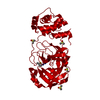
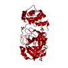
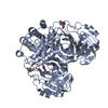
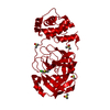
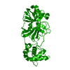
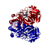
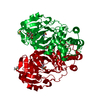
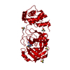
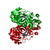
[English] 日本語
Yorodumi- PDB-5rgt: PanDDA analysis group deposition SARS-CoV-2 main protease fragmen... -
+Open data
-Basic information
Entry | Database: PDB / ID: 5rgt | ||||||
---|---|---|---|---|---|---|---|
Title | PanDDA analysis group deposition SARS-CoV-2 main protease fragment screen -- Crystal Structure of SARS-CoV-2 main protease in complex with Z4439011607 (Mpro-x2540) | ||||||
Components | 3C-like proteinase | ||||||
Keywords | HYDROLASE/HYDROLASE INHIBITOR / SGC - Diamond I04-1 fragment screening / PanDDA / XChemExplorer / HYDROLASE-HYDROLASE INHIBITOR complex | ||||||
Function / homology | Function and homology information protein guanylyltransferase activity / RNA endonuclease activity, producing 3'-phosphomonoesters / mRNA guanylyltransferase activity / 5'-3' RNA helicase activity / Lyases; Phosphorus-oxygen lyases / symbiont-mediated suppression of host cytoplasmic pattern recognition receptor signaling pathway via inhibition of TBK1 activity / Assembly of the SARS-CoV-2 Replication-Transcription Complex (RTC) / Maturation of replicase proteins / ISG15-specific peptidase activity / Transcription of SARS-CoV-2 sgRNAs ...protein guanylyltransferase activity / RNA endonuclease activity, producing 3'-phosphomonoesters / mRNA guanylyltransferase activity / 5'-3' RNA helicase activity / Lyases; Phosphorus-oxygen lyases / symbiont-mediated suppression of host cytoplasmic pattern recognition receptor signaling pathway via inhibition of TBK1 activity / Assembly of the SARS-CoV-2 Replication-Transcription Complex (RTC) / Maturation of replicase proteins / ISG15-specific peptidase activity / Transcription of SARS-CoV-2 sgRNAs / Translation of Replicase and Assembly of the Replication Transcription Complex / TRAF3-dependent IRF activation pathway / Replication of the SARS-CoV-2 genome / snRNP Assembly / double membrane vesicle viral factory outer membrane / Hydrolases; Acting on ester bonds; Exoribonucleases producing 5'-phosphomonoesters / 5'-3' DNA helicase activity / SARS coronavirus main proteinase / 3'-5'-RNA exonuclease activity / host cell endoplasmic reticulum-Golgi intermediate compartment / host cell endosome / symbiont-mediated suppression of host toll-like receptor signaling pathway / symbiont-mediated degradation of host mRNA / mRNA guanylyltransferase / symbiont-mediated suppression of host ISG15-protein conjugation / G-quadruplex RNA binding / omega peptidase activity / symbiont-mediated suppression of host cytoplasmic pattern recognition receptor signaling pathway via inhibition of IRF3 activity / SARS-CoV-2 modulates host translation machinery / mRNA (guanine-N7)-methyltransferase / host cell Golgi apparatus / methyltransferase cap1 / symbiont-mediated perturbation of host ubiquitin-like protein modification / DNA helicase / mRNA (nucleoside-2'-O-)-methyltransferase activity / mRNA 5'-cap (guanine-N7-)-methyltransferase activity / ubiquitinyl hydrolase 1 / cysteine-type deubiquitinase activity / Hydrolases; Acting on peptide bonds (peptidases); Cysteine endopeptidases / single-stranded RNA binding / host cell perinuclear region of cytoplasm / host cell endoplasmic reticulum membrane / viral protein processing / lyase activity / symbiont-mediated suppression of host type I interferon-mediated signaling pathway / RNA helicase / induction by virus of host autophagy / copper ion binding / cysteine-type endopeptidase activity / RNA-directed RNA polymerase / viral RNA genome replication / virus-mediated perturbation of host defense response / RNA-dependent RNA polymerase activity / DNA-templated transcription / lipid binding / host cell nucleus / SARS-CoV-2 activates/modulates innate and adaptive immune responses / ATP hydrolysis activity / proteolysis / RNA binding / zinc ion binding / ATP binding / membrane Similarity search - Function | ||||||
Biological species | Severe acute respiratory syndrome coronavirus 2 | ||||||
Method | X-RAY DIFFRACTION / SYNCHROTRON / FOURIER SYNTHESIS / molecular replacement / Resolution: 2.22 Å | ||||||
Authors | Fearon, D. / Owen, C.D. / Douangamath, A. / Lukacik, P. / Powell, A.J. / Strain-Damerell, C.M. / Resnick, E. / Krojer, T. / Gehrtz, P. / Wild, C. ...Fearon, D. / Owen, C.D. / Douangamath, A. / Lukacik, P. / Powell, A.J. / Strain-Damerell, C.M. / Resnick, E. / Krojer, T. / Gehrtz, P. / Wild, C. / Aimon, A. / Brandao-Neto, J. / Carbery, A. / Dunnett, L. / Skyner, R. / Snee, M. / London, N. / Walsh, M.A. / von Delft, F. | ||||||
Citation | Journal: To Be Published Title: PanDDA analysis group deposition SARS-CoV-2 main protease fragment screen Authors: Fearon, D. / Owen, C.D. / Douangamath, A. / Lukacik, P. / Powell, A.J. / Strain-Damerell, C.M. / Resnick, E. / Krojer, T. / Gehrtz, P. / Wild, C. / Aimon, A. / Brandao-Neto, J. / Carbery, A. ...Authors: Fearon, D. / Owen, C.D. / Douangamath, A. / Lukacik, P. / Powell, A.J. / Strain-Damerell, C.M. / Resnick, E. / Krojer, T. / Gehrtz, P. / Wild, C. / Aimon, A. / Brandao-Neto, J. / Carbery, A. / Dunnett, L. / Skyner, R. / Snee, M. / London, N. / Walsh, M.A. / von Delft, F. | ||||||
History |
|
-Structure visualization
Structure viewer | Molecule: MolmilJmol/JSmol |
---|
-Downloads & links
-Download
PDBx/mmCIF format | 5rgt.cif.gz | 72.6 KB | Display | PDBx/mmCIF format |
---|---|---|---|---|
PDB format | pdb5rgt.ent.gz | 56.8 KB | Display | PDB format |
PDBx/mmJSON format | 5rgt.json.gz | Tree view | PDBx/mmJSON format | |
Others | Other downloads |
-Validation report
Summary document | 5rgt_validation.pdf.gz | 765.5 KB | Display | wwPDB validaton report |
---|---|---|---|---|
Full document | 5rgt_full_validation.pdf.gz | 766.5 KB | Display | |
Data in XML | 5rgt_validation.xml.gz | 14.9 KB | Display | |
Data in CIF | 5rgt_validation.cif.gz | 21 KB | Display | |
Arichive directory | https://data.pdbj.org/pub/pdb/validation_reports/rg/5rgt ftp://data.pdbj.org/pub/pdb/validation_reports/rg/5rgt | HTTPS FTP |
-Group deposition
ID | G_1002155 (18 entries) |
---|---|
Title | PanDDA analysis group deposition SARS-CoV-2 main protease fragment screen |
Type | changed state |
Description | SARS-CoV-2 main protease from Coronaviridae sp. screened against COVID Moonshot compounds by X-ray Crystallography at the XChem facility of Diamond Light Source beamline I04-1 |
-Related structure data
Related structure data | 6lu7S S: Starting model for refinement |
---|---|
Similar structure data |
-Links
-Assembly
Deposited unit |
| ||||||||
---|---|---|---|---|---|---|---|---|---|
1 |
| ||||||||
Unit cell |
|
-Components
#1: Protein | Mass: 33825.547 Da / Num. of mol.: 1 Source method: isolated from a genetically manipulated source Source: (gene. exp.) Severe acute respiratory syndrome coronavirus 2 Gene: rep, 1a-1b / Production host: Escherichia coli (E. coli) References: UniProt: P0DTD1, SARS coronavirus main proteinase | ||||||
---|---|---|---|---|---|---|---|
#2: Chemical | ChemComp-DMS / #3: Chemical | ChemComp-UHS / | #4: Water | ChemComp-HOH / | Has ligand of interest | Y | |
-Experimental details
-Experiment
Experiment | Method: X-RAY DIFFRACTION / Number of used crystals: 1 |
---|
-Sample preparation
Crystal | Density Matthews: 1.91 Å3/Da / Density % sol: 35.71 % / Mosaicity: 0.33 ° |
---|---|
Crystal grow | Temperature: 293.15 K / Method: vapor diffusion, sitting drop / pH: 6.5 / Details: 15% PEG 4K, 5% DMSO, 0.1M MES pH 6.5 |
-Data collection
Diffraction | Mean temperature: 100 K | ||||||||||||||||||||||||||||||
---|---|---|---|---|---|---|---|---|---|---|---|---|---|---|---|---|---|---|---|---|---|---|---|---|---|---|---|---|---|---|---|
Diffraction source | Source: SYNCHROTRON / Site: Diamond / Beamline: I04-1 / Wavelength: 0.913 Å | ||||||||||||||||||||||||||||||
Detector | Type: DECTRIS PILATUS 6M / Detector: PIXEL / Date: Apr 9, 2020 | ||||||||||||||||||||||||||||||
Radiation | Protocol: SINGLE WAVELENGTH / Scattering type: x-ray | ||||||||||||||||||||||||||||||
Radiation wavelength | Wavelength: 0.913 Å / Relative weight: 1 | ||||||||||||||||||||||||||||||
Reflection | Resolution: 2.22→47.55 Å / Num. obs: 12786 / % possible obs: 99.8 % / Redundancy: 3.8 % / CC1/2: 0.989 / Rmerge(I) obs: 0.196 / Rpim(I) all: 0.116 / Rrim(I) all: 0.228 / Net I/σ(I): 5.2 / Num. measured all: 48314 / Scaling rejects: 0 | ||||||||||||||||||||||||||||||
Reflection shell | Diffraction-ID: 1
|
-Phasing
Phasing | Method: molecular replacement |
---|
-Processing
Software |
| |||||||||||||||||||||||||||||||||||||||||||||||||||||||||||||||||||||||||||
---|---|---|---|---|---|---|---|---|---|---|---|---|---|---|---|---|---|---|---|---|---|---|---|---|---|---|---|---|---|---|---|---|---|---|---|---|---|---|---|---|---|---|---|---|---|---|---|---|---|---|---|---|---|---|---|---|---|---|---|---|---|---|---|---|---|---|---|---|---|---|---|---|---|---|---|---|
Refinement | Method to determine structure: FOURIER SYNTHESIS Starting model: 6LU7 Resolution: 2.22→47.59 Å / Cor.coef. Fo:Fc: 0.94 / Cor.coef. Fo:Fc free: 0.915 / SU B: 13.185 / SU ML: 0.291 / Cross valid method: THROUGHOUT / σ(F): 0 / ESU R: 0.604 / ESU R Free: 0.282 / Stereochemistry target values: MAXIMUM LIKELIHOOD Details: HYDROGENS HAVE BEEN ADDED IN THE RIDING POSITIONS U VALUES : REFINED INDIVIDUALLY
| |||||||||||||||||||||||||||||||||||||||||||||||||||||||||||||||||||||||||||
Solvent computation | Ion probe radii: 0.8 Å / Shrinkage radii: 0.8 Å / VDW probe radii: 1.2 Å / Solvent model: MASK | |||||||||||||||||||||||||||||||||||||||||||||||||||||||||||||||||||||||||||
Displacement parameters | Biso max: 84 Å2 / Biso mean: 37.133 Å2 / Biso min: 23.34 Å2
| |||||||||||||||||||||||||||||||||||||||||||||||||||||||||||||||||||||||||||
Refinement step | Cycle: final / Resolution: 2.22→47.59 Å
| |||||||||||||||||||||||||||||||||||||||||||||||||||||||||||||||||||||||||||
Refine LS restraints |
| |||||||||||||||||||||||||||||||||||||||||||||||||||||||||||||||||||||||||||
LS refinement shell | Resolution: 2.218→2.276 Å / Total num. of bins used: 20
|