 Movie
Movie Controller

[English] 日本語
 Yorodumi
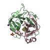
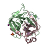
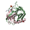
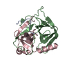
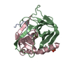
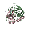
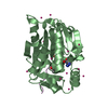
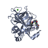
- PDB-5r46: Crystal Structure of deuterated gamma-Chymotrypsin at pH 5.6, roo... -
+ Open data

- Basic information
| Entry | Database: PDB / ID: 5r46 | |||||||||
|---|---|---|---|---|---|---|---|---|---|---|
| Title | Crystal Structure of deuterated gamma-Chymotrypsin at pH 5.6, room temperature | |||||||||
 Components Components |
| |||||||||
 Keywords Keywords | HYDROLASE / serine protease / hydrolase-peptide complex | |||||||||
| Function / homology |  Function and homology information Function and homology informationchymotrypsin / serpin family protein binding / serine protease inhibitor complex / digestion / serine-type endopeptidase activity / proteolysis / extracellular region Similarity search - Function | |||||||||
| Biological species |  | |||||||||
| Method |  X-RAY DIFFRACTION / SYNCHROTRON / FOURIER SYNTHESIS / Resolution: 1.05 Å X-RAY DIFFRACTION / SYNCHROTRON / FOURIER SYNTHESIS / Resolution: 1.05 Å | |||||||||
 Authors Authors | Kreinbring, C.A. / Wilson, M.A. / Kovalevsky, A.Y. / Blakeley, M.P. / Fisher, S.Z. / Lazar, L.M. / Moulin, A.G. / Novak, W.R. / Petsko, G.A. / Ringe, D. | |||||||||
| Funding support |  United States, 2items United States, 2items
| |||||||||
 Citation Citation | Journal: To be published Title: Effect of Temperature and pH on Ionizable Residues in gamma-Chymotrypsin: a X-ray and Neutron Crystallography Study Authors: Kreinbring, C.A. / Wilson, M.A. / Kovalevsky, A.Y. / Blakeley, M.P. / Fisher, S.Z. / Lazar, L.M. / Moulin, A.G. / Novak, W.R. / Petsko, G.A. / Ringe, D. | |||||||||
| History |
|
- Structure visualization
| Structure viewer | Molecule: MolmilJmol/JSmol |
|---|
- Downloads & links
-Download
| PDBx/mmCIF format | 5r46.cif.gz | 115.8 KB | Display | PDBx/mmCIF format |
|---|---|---|---|---|
| PDB format | pdb5r46.ent.gz | 89.7 KB | Display | PDB format |
| PDBx/mmJSON format | 5r46.json.gz | Tree view | PDBx/mmJSON format | |
| Others |  Other downloads Other downloads |
-Validation report
| Arichive directory | https://data.pdbj.org/pub/pdb/validation_reports/r4/5r46ftp://data.pdbj.org/pub/pdb/validation_reports/r4/5r46 | HTTPS FTP |
|---|
-Group deposition
| ID | G_1002095 (4 entries) |
|---|---|
| Title | Effect of Temperature and pH on Ionizable Residues in gamma-Chymotrypsin: a X-ray and Neutron Crystallography Study |
| Type | undefined |
| Description | gamma-Chymotrypsin at pH 5.6 |


-Related structure data
| Related structure data |  1gctS S: Starting model for refinement |
|---|---|
| Similar structure data |

-Links
 PDBj
PDBj

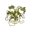
- Assembly
| Deposited unit | 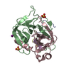
| |||||||||
|---|---|---|---|---|---|---|---|---|---|---|
| 1 |
| |||||||||
| Unit cell |
| |||||||||
| Components on special symmetry positions |
|
-Components
-Protein/peptide , 3 types, 3 molecules ADE
| #1: Protein/peptide | Mass: 1253.511 Da / Num. of mol.: 1 / Source method: isolated from a natural source / Source: (natural) |
|---|---|
| #4: Protein/peptide | Mass: 574.627 Da / Num. of mol.: 1 / Source method: isolated from a natural source / Source: (natural) |
| #5: Protein/peptide | Mass: 535.590 Da / Num. of mol.: 1 / Source method: isolated from a natural source / Source: (natural) |
-Protein , 2 types, 2 molecules BC
| #2: Protein | Mass: 13934.556 Da / Num. of mol.: 1 / Source method: isolated from a natural source / Source: (natural) |
|---|---|
| #3: Protein | Mass: 10074.495 Da / Num. of mol.: 1 / Source method: isolated from a natural source / Source: (natural) |
-Non-polymers , 3 types, 168 molecules 


| #6: Chemical | ChemComp-IOD /  Mass: 126.904 Da / Num. of mol.: 1 / Source method: obtained synthetically / Formula: I Mass: 126.904 Da / Num. of mol.: 1 / Source method: obtained synthetically / Formula: I | ||
|---|---|---|---|
| #7: Chemical |  Mass: 96.063 Da / Num. of mol.: 2 / Source method: obtained synthetically / Formula: SO4 Mass: 96.063 Da / Num. of mol.: 2 / Source method: obtained synthetically / Formula: SO4#8: Water | ChemComp-HOH / | Mass: 18.015 Da / Num. of mol.: 165 / Source method: isolated from a natural source / Formula: H2O |
-Details
| Has protein modification | Y |
|---|
-Experimental details
-Experiment
| Experiment | Method: X-RAY DIFFRACTION / Number of used crystals: 1 |
|---|
- Sample preparation
| Crystal | Density Matthews: 2.24 Å3/Da / Density % sol: 45.02 % |
|---|---|
| Crystal grow | Temperature: 297 K / Method: vapor diffusion, sitting drop Details: 45% saturated ammonium sulfate, 0.75% saturated cetyltrimethylammonium bromide, 100 mM sodium iodide; sodium malonate PH range: 10 mM sodium cacodylate pH 6.0 |
-Data collection
| Diffraction | Mean temperature: 297 K | |||||||||||||||||||||||||||||||||||||||||||||||||||||||||||||||||||||||||||||
|---|---|---|---|---|---|---|---|---|---|---|---|---|---|---|---|---|---|---|---|---|---|---|---|---|---|---|---|---|---|---|---|---|---|---|---|---|---|---|---|---|---|---|---|---|---|---|---|---|---|---|---|---|---|---|---|---|---|---|---|---|---|---|---|---|---|---|---|---|---|---|---|---|---|---|---|---|---|---|
| Diffraction source | Source: SYNCHROTRON / Site: APS / Beamline: 23-ID-B / Wavelength: 0.9 Å | |||||||||||||||||||||||||||||||||||||||||||||||||||||||||||||||||||||||||||||
| Detector | Type: MARMOSAIC 300 mm CCD / Detector: CCD / Date: Jul 15, 2009 | |||||||||||||||||||||||||||||||||||||||||||||||||||||||||||||||||||||||||||||
| Radiation | Protocol: SINGLE WAVELENGTH / Monochromatic (M) / Laue (L): M / Scattering type: x-ray | |||||||||||||||||||||||||||||||||||||||||||||||||||||||||||||||||||||||||||||
| Radiation wavelength | Wavelength: 0.9 Å / Relative weight: 1 | |||||||||||||||||||||||||||||||||||||||||||||||||||||||||||||||||||||||||||||
| Reflection | Resolution: 1.05→50 Å / Num. obs: 111717 / % possible obs: 99.6 % / Redundancy: 39.3 % / Rmerge(I) obs: 0.109 / Χ2: 1.125 / Net I/σ(I): 13.1 / Num. measured all: 4389000 | |||||||||||||||||||||||||||||||||||||||||||||||||||||||||||||||||||||||||||||
| Reflection shell |
|
- Processing
| Software |
| |||||||||||||||||||||||||||||||||||||||||||||||||||||||||||||||||||||||||||
|---|---|---|---|---|---|---|---|---|---|---|---|---|---|---|---|---|---|---|---|---|---|---|---|---|---|---|---|---|---|---|---|---|---|---|---|---|---|---|---|---|---|---|---|---|---|---|---|---|---|---|---|---|---|---|---|---|---|---|---|---|---|---|---|---|---|---|---|---|---|---|---|---|---|---|---|---|
| Refinement | Method to determine structure: FOURIER SYNTHESIS Starting model: 1GCT Resolution: 1.05→56.64 Å / Cor.coef. Fo:Fc: 0.984 / Cor.coef. Fo:Fc free: 0.982 / SU B: 0.484 / SU ML: 0.011 / Cross valid method: THROUGHOUT / σ(F): 0 / ESU R: 0.019 / ESU R Free: 0.018 / Stereochemistry target values: MAXIMUM LIKELIHOOD Details: HYDROGENS HAVE BEEN ADDED IN THE RIDING POSITIONS U VALUES : REFINED INDIVIDUALLY
| |||||||||||||||||||||||||||||||||||||||||||||||||||||||||||||||||||||||||||
| Solvent computation | Ion probe radii: 0.8 Å / Shrinkage radii: 0.8 Å / VDW probe radii: 1.2 Å / Solvent model: MASK | |||||||||||||||||||||||||||||||||||||||||||||||||||||||||||||||||||||||||||
| Displacement parameters | Biso max: 107.66 Å2 / Biso mean: 17.772 Å2 / Biso min: 8.97 Å2
| |||||||||||||||||||||||||||||||||||||||||||||||||||||||||||||||||||||||||||
| Refinement step | Cycle: final / Resolution: 1.05→56.64 Å
| |||||||||||||||||||||||||||||||||||||||||||||||||||||||||||||||||||||||||||
| Refine LS restraints |
| |||||||||||||||||||||||||||||||||||||||||||||||||||||||||||||||||||||||||||
| LS refinement shell | Resolution: 1.05→1.077 Å / Rfactor Rfree error: 0 / Total num. of bins used: 20
|
