regulation of Fas signaling pathway / regulation of viral process / maintenance of protein location / chromo shadow domain binding / response to type I interferon / negative regulation of protein export from nucleus / negative regulation of viral transcription / regulation of extrinsic apoptotic signaling pathway via death domain receptors / negative regulation of endothelial cell migration / type I interferon-mediated signaling pathway ...regulation of Fas signaling pathway / regulation of viral process / maintenance of protein location / chromo shadow domain binding / response to type I interferon / negative regulation of protein export from nucleus / negative regulation of viral transcription / regulation of extrinsic apoptotic signaling pathway via death domain receptors / negative regulation of endothelial cell migration / type I interferon-mediated signaling pathway / regulation of angiogenesis / response to retinoic acid / type II interferon-mediated signaling pathway / retinoic acid receptor signaling pathway / SUMOylation of DNA damage response and repair proteins / response to type II interferon / response to cytokine / nuclear periphery / telomere maintenance / DNA damage response, signal transduction by p53 class mediator / PML body / kinase binding / Interferon gamma signaling / transcription corepressor activity / DNA-binding transcription activator activity, RNA polymerase II-specific / RNA polymerase II-specific DNA-binding transcription factor binding / DNA-binding transcription factor activity, RNA polymerase II-specific / chromosome, telomeric region / protein dimerization activity / nuclear body / protein domain specific binding / negative regulation of DNA-templated transcription / regulation of transcription by RNA polymerase II / nucleolus / negative regulation of transcription by RNA polymerase II / protein homodimerization activity / positive regulation of transcription by RNA polymerase II / DNA binding / nucleoplasm / identical protein binding / nucleus / cytoplasm Similarity search - Function
HSR domain / Nuclear body protein Sp110/Sp140/Sp140L / HSR domain / HSR domain profile. / SAND domain / SAND domain / SAND domain profile. / SAND domain / SAND-like domain superfamily / HMG-box domain ...HSR domain / Nuclear body protein Sp110/Sp140/Sp140L / HSR domain / HSR domain profile. / SAND domain / SAND domain / SAND domain profile. / SAND domain / SAND-like domain superfamily / HMG-box domain / HMG (high mobility group) box / HMG boxes A and B DNA-binding domains profile. / high mobility group / High mobility group box domain / High mobility group box domain superfamily / Zinc/RING finger domain, C3HC4 (zinc finger) / Herpes Virus-1 / Bromodomain-like / Histone Acetyltransferase; Chain A / Up-down Bundle / 2-Layer Sandwich / Mainly Alpha / Alpha Beta Similarity search - Domain/homology
PanDDA analysis group deposition of models of ground state datasets
Type
ground state
Description
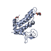
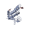
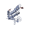
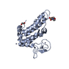
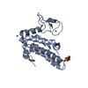
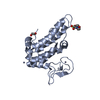
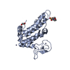
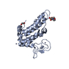
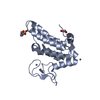
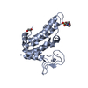
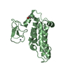
human SP100 screened against the Maybridge Library by X-ray Crystallography at the XChem facility of Diamond Light Source beamline I04-1. Check out the PanDDA event maps at https://zenodo.org/record/290201/files/0_index.html
In the structure databanks used in Yorodumi, some data are registered as the other names, "COVID-19 virus" and "2019-nCoV". Here are the details of the virus and the list of structure data.
Jan 31, 2019. EMDB accession codes are about to change! (news from PDBe EMDB page)
EMDB accession codes are about to change! (news from PDBe EMDB page)
The allocation of 4 digits for EMDB accession codes will soon come to an end. Whilst these codes will remain in use, new EMDB accession codes will include an additional digit and will expand incrementally as the available range of codes is exhausted. The current 4-digit format prefixed with “EMD-” (i.e. EMD-XXXX) will advance to a 5-digit format (i.e. EMD-XXXXX), and so on. It is currently estimated that the 4-digit codes will be depleted around Spring 2019, at which point the 5-digit format will come into force.
The EM Navigator/Yorodumi systems omit the EMD- prefix.
Related info.:Q: What is EMD? / ID/Accession-code notation in Yorodumi/EM Navigator
Yorodumi is a browser for structure data from EMDB, PDB, SASBDB, etc.
This page is also the successor to EM Navigator detail page, and also detail information page/front-end page for Omokage search.
The word "yorodu" (or yorozu) is an old Japanese word meaning "ten thousand". "mi" (miru) is to see.
Related info.:EMDB / PDB / SASBDB / Comparison of 3 databanks / Yorodumi Search / Aug 31, 2016. New EM Navigator & Yorodumi / Yorodumi Papers / Jmol/JSmol / Function and homology information / Changes in new EM Navigator and Yorodumi
 Movie
Movie Controller

 Yorodumi
 Open data
 Basic information
 Components
 Keywords
 Function and homology information
 Homo sapiens (human)
 X-RAY DIFFRACTION /
  Authors
 Citation
 Structure visualization
 Downloads & links
 Other downloads


 PDBj
PDBj


 Assembly
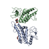


 Mass: 65.409 Da / Num. of mol.: 5 / Source method: obtained synthetically / Formula: Zn
 Mass: 195.237 Da / Num. of mol.: 1 / Source method: obtained synthetically / Formula: C6H13NO4S / Comment: pH buffer*YM
 Mass: 62.068 Da / Num. of mol.: 2 / Source method: obtained synthetically / Formula: C2H6O2
 Mass: 18.015 Da / Num. of mol.: 579 / Source method: isolated from a natural source / Formula: H2O
 Sample preparation
 / Beamline: I04-1 / Wavelength: 0.92 Å
 Processing