regulation of Fas signaling pathway / regulation of viral process / maintenance of protein location / chromo shadow domain binding / response to type I interferon / negative regulation of protein export from nucleus / negative regulation of viral transcription / regulation of extrinsic apoptotic signaling pathway via death domain receptors / negative regulation of endothelial cell migration / type I interferon-mediated signaling pathway ...regulation of Fas signaling pathway / regulation of viral process / maintenance of protein location / chromo shadow domain binding / response to type I interferon / negative regulation of protein export from nucleus / negative regulation of viral transcription / regulation of extrinsic apoptotic signaling pathway via death domain receptors / negative regulation of endothelial cell migration / type I interferon-mediated signaling pathway / regulation of angiogenesis / response to retinoic acid / type II interferon-mediated signaling pathway / response to type II interferon / retinoic acid receptor signaling pathway / SUMOylation of DNA damage response and repair proteins / response to cytokine / nuclear periphery / telomere maintenance / DNA damage response, signal transduction by p53 class mediator / PML body / kinase binding / Interferon gamma signaling / transcription corepressor activity / DNA-binding transcription activator activity, RNA polymerase II-specific / RNA polymerase II-specific DNA-binding transcription factor binding / DNA-binding transcription factor activity, RNA polymerase II-specific / chromosome, telomeric region / protein dimerization activity / nuclear body / protein domain specific binding / negative regulation of DNA-templated transcription / regulation of transcription by RNA polymerase II / nucleolus / negative regulation of transcription by RNA polymerase II / protein homodimerization activity / positive regulation of transcription by RNA polymerase II / DNA binding / zinc ion binding / nucleoplasm / identical protein binding / nucleus / cytoplasm Similarity search - Function
Nuclear body protein Sp140 / HSR domain / Nuclear body protein Sp110/Sp140/Sp140L / HSR domain / HSR domain profile. / SAND domain / SAND domain / SAND domain profile. / SAND domain / SAND-like domain superfamily ...Nuclear body protein Sp140 / HSR domain / Nuclear body protein Sp110/Sp140/Sp140L / HSR domain / HSR domain profile. / SAND domain / SAND domain / SAND domain profile. / SAND domain / SAND-like domain superfamily / HMG-box domain / HMG (high mobility group) box / HMG boxes A and B DNA-binding domains profile. / high mobility group / High mobility group box domain / High mobility group box domain superfamily / Zinc/RING finger domain, C3HC4 (zinc finger) / Herpes Virus-1 / Zinc finger, PHD-type, conserved site / Zinc finger PHD-type signature. / Zinc finger PHD-type profile. / Zinc finger, PHD-finger / Zinc finger, PHD-type / PHD zinc finger / Bromodomain-like / Histone Acetyltransferase; Chain A / Zinc finger, FYVE/PHD-type / Bromodomain / bromo domain / Bromodomain / Bromodomain-like superfamily / Zinc finger, RING/FYVE/PHD-type / Up-down Bundle / 2-Layer Sandwich / Mainly Alpha / Alpha Beta Similarity search - Domain/homology
Method to determine structure: SAD / Resolution: 1.6→27.72 Å / Cor.coef. Fo:Fc: 0.962 / Cor.coef. Fo:Fc free: 0.955 / SU B: 2.337 / SU ML: 0.043 / Cross valid method: THROUGHOUT / σ(F): 0 / ESU R: 0.078 / ESU R Free: 0.076 / Stereochemistry target values: MAXIMUM LIKELIHOOD / Details: HYDROGENS HAVE BEEN ADDED IN THE RIDING POSITIONS
Rfactor
Num. reflection
% reflection
Selection details
Rfree
0.19229
2950
5.1 %
RANDOM
Rwork
0.17303
-
-
-
all
0.17402
55205
-
-
obs
0.17402
55205
94.17 %
-
Solvent computation
Ion probe radii: 0.8 Å / Shrinkage radii: 0.8 Å / VDW probe radii: 1.2 Å / Solvent model: MASK
Displacement parameters
Biso mean: 25.806 Å2
Baniso -1
Baniso -2
Baniso -3
1-
-0.6 Å2
0 Å2
0.38 Å2
2-
-
-0.3 Å2
-0 Å2
3-
-
-
0.67 Å2
Refinement step
Cycle: LAST / Resolution: 1.6→27.72 Å
Protein
Nucleic acid
Ligand
Solvent
Total
Num. atoms
2866
0
32
200
3098
Refine LS restraints
Refine-ID
Type
Dev ideal
Dev ideal target
Number
X-RAY DIFFRACTION
r_bond_refined_d
0.011
0.019
3038
X-RAY DIFFRACTION
r_bond_other_d
0.001
0.02
2826
X-RAY DIFFRACTION
r_angle_refined_deg
1.392
1.939
4088
X-RAY DIFFRACTION
r_angle_other_deg
0.804
3
6520
X-RAY DIFFRACTION
r_dihedral_angle_1_deg
5.27
5
359
X-RAY DIFFRACTION
r_dihedral_angle_2_deg
34.802
23.72
164
X-RAY DIFFRACTION
r_dihedral_angle_3_deg
12.542
15
557
X-RAY DIFFRACTION
r_dihedral_angle_4_deg
14.49
15
25
X-RAY DIFFRACTION
r_chiral_restr
0.083
0.2
416
X-RAY DIFFRACTION
r_gen_planes_refined
0.007
0.021
3442
X-RAY DIFFRACTION
r_gen_planes_other
0.001
0.02
775
X-RAY DIFFRACTION
r_mcbond_it
1.564
1.914
1409
X-RAY DIFFRACTION
r_mcbond_other
1.556
1.912
1408
X-RAY DIFFRACTION
r_mcangle_it
2.504
2.856
1765
X-RAY DIFFRACTION
r_mcangle_other
2.505
2.858
1766
X-RAY DIFFRACTION
r_scbond_it
2.59
2.261
1629
X-RAY DIFFRACTION
r_scbond_other
2.589
2.261
1629
X-RAY DIFFRACTION
r_scangle_other
4.127
3.27
2320
X-RAY DIFFRACTION
r_long_range_B_refined
5.732
15.906
3624
X-RAY DIFFRACTION
r_long_range_B_other
5.689
15.541
3527
LS refinement shell
Resolution: 1.598→1.639 Å / Total num. of bins used: 20
Rfactor
Num. reflection
% reflection
Rfree
0.18
212
-
Rwork
0.184
3848
-
obs
-
-
89.55 %
Refinement TLS params.
Method: refined / Refine-ID: X-RAY DIFFRACTION
ID
L11 (°2)
L12 (°2)
L13 (°2)
L22 (°2)
L23 (°2)
L33 (°2)
S11 (Å °)
S12 (Å °)
S13 (Å °)
S21 (Å °)
S22 (Å °)
S23 (Å °)
S31 (Å °)
S32 (Å °)
S33 (Å °)
T11 (Å2)
T12 (Å2)
T13 (Å2)
T22 (Å2)
T23 (Å2)
T33 (Å2)
Origin x (Å)
Origin y (Å)
Origin z (Å)
1
0.3951
-0.2914
-0.0312
0.6138
-0.0411
0.2724
0.0726
0.0495
0.0044
-0.0094
-0.0698
0.0003
0.0251
-0.0093
-0.0028
0.0408
0.0187
-0.0034
0.0237
0.0007
0.0157
26.1287
3.8545
16.319
2
0.746
-0.0057
0.2432
0.2128
0.109
0.3342
0.11
0.0411
0.0471
-0.0274
-0.0722
-0.0682
-0.0153
-0.0703
-0.0377
0.0326
0.0344
0.0152
0.0565
0.0229
0.0283
-2.2987
24.0641
19.3789
Refinement TLS group
ID
Refine-ID
Refine TLS-ID
Auth asym-ID
Auth seq-ID
1
X-RAY DIFFRACTION
1
A
701 - 878
2
X-RAY DIFFRACTION
2
B
701 - 875
+
About Yorodumi
-
News
-
Feb 9, 2022. New format data for meta-information of EMDB entries
New format data for meta-information of EMDB entries
Version 3 of the EMDB header file is now the official format.
The previous official version 1.9 will be removed from the archive.
In the structure databanks used in Yorodumi, some data are registered as the other names, "COVID-19 virus" and "2019-nCoV". Here are the details of the virus and the list of structure data.
Jan 31, 2019. EMDB accession codes are about to change! (news from PDBe EMDB page)
EMDB accession codes are about to change! (news from PDBe EMDB page)
The allocation of 4 digits for EMDB accession codes will soon come to an end. Whilst these codes will remain in use, new EMDB accession codes will include an additional digit and will expand incrementally as the available range of codes is exhausted. The current 4-digit format prefixed with “EMD-” (i.e. EMD-XXXX) will advance to a 5-digit format (i.e. EMD-XXXXX), and so on. It is currently estimated that the 4-digit codes will be depleted around Spring 2019, at which point the 5-digit format will come into force.
The EM Navigator/Yorodumi systems omit the EMD- prefix.
Related info.:Q: What is EMD? / ID/Accession-code notation in Yorodumi/EM Navigator
Yorodumi is a browser for structure data from EMDB, PDB, SASBDB, etc.
This page is also the successor to EM Navigator detail page, and also detail information page/front-end page for Omokage search.
The word "yorodu" (or yorozu) is an old Japanese word meaning "ten thousand". "mi" (miru) is to see.
Related info.:EMDB / PDB / SASBDB / Comparison of 3 databanks / Yorodumi Search / Aug 31, 2016. New EM Navigator & Yorodumi / Yorodumi Papers / Jmol/JSmol / Function and homology information / Changes in new EM Navigator and Yorodumi
 Movie
Movie Controller

 Yorodumi
 Open data
 Basic information
 Components
 Keywords
 Function and homology information
 Homo sapiens (human)
 X-RAY DIFFRACTION /
  Authors
 Citation
 Structure visualization
 Downloads & links
 Other downloads


 PDBj
PDBj


 Assembly
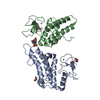

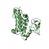


 Mass: 65.409 Da / Num. of mol.: 4 / Source method: obtained synthetically / Formula: Zn
 Mass: 195.237 Da / Num. of mol.: 1 / Source method: obtained synthetically / Formula: C6H13NO4S / Comment: pH buffer*YM
 Mass: 62.068 Da / Num. of mol.: 4 / Source method: obtained synthetically / Formula: C2H6O2
 Mass: 18.015 Da / Num. of mol.: 200 / Source method: isolated from a natural source / Formula: H2O
 Sample preparation
 / Beamline: I04 / Wavelength: 1.282 Å
 Processing