 Movie
Movie Controller


+ Open data

- Basic information
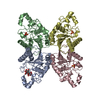
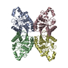
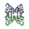
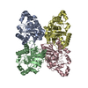
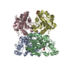
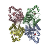
| Entry | Database: PDB / ID: 5pgm | ||||||
|---|---|---|---|---|---|---|---|
| Title | SACCHAROMYCES CEREVISIAE PHOSPHOGLYCERATE MUTASE | ||||||
 Components Components | PHOSPHOGLYCERATE MUTASE 1 | ||||||
 Keywords Keywords | ISOMERASE / TRANSFERASE (PHOSPHORYL) / GLYCOLYTIC ENZYME | ||||||
| Function / homology |  Function and homology information Function and homology informationphosphoglycerate mutase (2,3-diphosphoglycerate-dependent) / phosphoglycerate mutase activity / canonical glycolysis / gluconeogenesis / glycolytic process / mitochondrial intermembrane space / mitochondrial outer membrane / mitochondrion / cytosol Similarity search - Function | ||||||
| Biological species |  | ||||||
| Method |  X-RAY DIFFRACTION / SYNCHROTRON / MOLECULAR REPLACEMENT / Resolution: 2.12 Å X-RAY DIFFRACTION / SYNCHROTRON / MOLECULAR REPLACEMENT / Resolution: 2.12 Å | ||||||
 Authors Authors | Rigden, D.J. / Phillips, S.E.V. / Fothergill-Gilmore, L.A. | ||||||
 Citation Citation | Journal: J.Mol.Biol. / Year: 1999 Title: Sulphate ions observed in the 2.12 A structure of a new crystal form of S. cerevisiae phosphoglycerate mutase provide insights into understanding the catalytic mechanism. Authors: Rigden, D.J. / Walter, R.A. / Phillips, S.E. / Fothergill-Gilmore, L.A. #1: Journal: J.Mol.Biol. / Year: 1998Title: The 2.3 A X-Ray Crystal Structure of S. Cerevisiae Phosphoglycerate Mutase Authors: Rigden, D.J. / Alexeev, D. / Phillips, S.E. / Fothergill-Gilmore, L.A. #2: Journal: Nature / Year: 1974Title: Structure of Yeast Phosphoglycerate Mutase Authors: Campbell, J.W. / Watson, H.C. / Hodgson, G.I. | ||||||
| History |
|
- Structure visualization
| Structure viewer | Molecule: MolmilJmol/JSmol |
|---|
- Downloads & links
-Download
| PDBx/mmCIF format | 5pgm.cif.gz | 411.2 KB | Display | PDBx/mmCIF format |
|---|---|---|---|---|
| PDB format | pdb5pgm.ent.gz | 337.1 KB | Display | PDB format |
| PDBx/mmJSON format | 5pgm.json.gz | Tree view | PDBx/mmJSON format | |
| Others |  Other downloads Other downloads |
-Validation report
| Arichive directory | https://data.pdbj.org/pub/pdb/validation_reports/pg/5pgmftp://data.pdbj.org/pub/pdb/validation_reports/pg/5pgm | HTTPS FTP |
|---|
-Related structure data
| Related structure data |  4pgmS S: Starting model for refinement |
|---|---|
| Similar structure data |


-Links
 PDBj
PDBj


- Assembly
| Deposited unit | 
| ||||||||
|---|---|---|---|---|---|---|---|---|---|
| 1 | 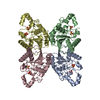
| ||||||||
| 2 | 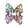
| ||||||||
| Unit cell |
| ||||||||
| Noncrystallographic symmetry (NCS) | NCS oper: (Code: given Matrix: (1, 0.00054, 0.00298), Vector: |
-Components
| #1: Protein | Mass: 27517.369 Da / Num. of mol.: 8 / Source method: isolated from a natural source / Source: (natural) #2: Chemical | ChemComp-SO4 /   Mass: 96.063 Da / Num. of mol.: 16 / Source method: obtained synthetically / Formula: SO4 Mass: 96.063 Da / Num. of mol.: 16 / Source method: obtained synthetically / Formula: SO4#3: Chemical | ChemComp-ALA / |   Type: L-peptide linking / Mass: 89.093 Da / Num. of mol.: 1 / Source method: obtained synthetically / Formula: C3H7NO2 Type: L-peptide linking / Mass: 89.093 Da / Num. of mol.: 1 / Source method: obtained synthetically / Formula: C3H7NO2#4: Water | ChemComp-HOH / |  Mass: 18.015 Da / Num. of mol.: 1638 / Source method: isolated from a natural source / Formula: H2O Mass: 18.015 Da / Num. of mol.: 1638 / Source method: isolated from a natural source / Formula: H2OHas protein modification | N | |
|---|
-Experimental details
-Experiment
| Experiment | Method: X-RAY DIFFRACTION / Number of used crystals: 1 |
|---|
- Sample preparation
| Crystal | Density Matthews: 2.89 Å3/Da / Density % sol: 57.1 % | ||||||||||||||||||||||||||||||||||||
|---|---|---|---|---|---|---|---|---|---|---|---|---|---|---|---|---|---|---|---|---|---|---|---|---|---|---|---|---|---|---|---|---|---|---|---|---|---|
| Crystal grow | pH: 8.65 / Details: pH 8.65 | ||||||||||||||||||||||||||||||||||||
| Crystal grow | *PLUS Method: vapor diffusion, sitting drop / Details: Rigden, D.J., (1998) J.Mol.Biol., 276, 449. | ||||||||||||||||||||||||||||||||||||
| Components of the solutions | *PLUS
|
-Data collection
| Diffraction | Mean temperature: 100 K |
|---|---|
| Diffraction source | Source: SYNCHROTRON / Site: SRS  / Beamline: PX9.5 / Wavelength: 0.95 / Beamline: PX9.5 / Wavelength: 0.95 |
| Detector | Type: MARRESEARCH / Detector: IMAGE PLATE / Date: Sep 1, 1997 / Details: TOROIDAL MIRROR |
| Radiation | Monochromator: SI(111) / Monochromatic (M) / Laue (L): M / Scattering type: x-ray |
| Radiation wavelength | Wavelength: 0.95 Å / Relative weight: 1 |
| Reflection | Resolution: 2.12→30 Å / Num. obs: 120182 / % possible obs: 89.5 % / Redundancy: 2.6 % / Rmerge(I) obs: 0.062 / Net I/σ(I): 8.3 |
| Reflection shell | Resolution: 2.12→2.24 Å / Redundancy: 2.4 % / Rmerge(I) obs: 0.182 / Mean I/σ(I) obs: 4 / % possible all: 80.5 |
| Reflection | *PLUS Lowest resolution: 30 Å / % possible obs: 90.8 % |
| Reflection shell | *PLUS Lowest resolution: 2.2 Å / % possible obs: 81.8 % / Rmerge(I) obs: 0.184 / Mean I/σ(I) obs: 3.9 |
- Processing
| Software |
| ||||||||||||||||||||||||||||||||||||||||||||||||||||||||||||
|---|---|---|---|---|---|---|---|---|---|---|---|---|---|---|---|---|---|---|---|---|---|---|---|---|---|---|---|---|---|---|---|---|---|---|---|---|---|---|---|---|---|---|---|---|---|---|---|---|---|---|---|---|---|---|---|---|---|---|---|---|---|
| Refinement | Method to determine structure: MOLECULAR REPLACEMENT Starting model: PDB ENTRY 4PGM Resolution: 2.12→30 Å / Rfactor Rfree error: 0.003 / Data cutoff high absF: 100000 / Data cutoff low absF: 0.001 / Cross valid method: THROUGHOUT / σ(F): 0
| ||||||||||||||||||||||||||||||||||||||||||||||||||||||||||||
| Displacement parameters | Biso mean: 23.4 Å2
| ||||||||||||||||||||||||||||||||||||||||||||||||||||||||||||
| Refine analyze | Luzzati d res low obs: 30 Å | ||||||||||||||||||||||||||||||||||||||||||||||||||||||||||||
| Refinement step | Cycle: LAST / Resolution: 2.12→30 Å
| ||||||||||||||||||||||||||||||||||||||||||||||||||||||||||||
| Refine LS restraints |
| ||||||||||||||||||||||||||||||||||||||||||||||||||||||||||||
| Refine LS restraints NCS | NCS model details: CONSTRAINTS | ||||||||||||||||||||||||||||||||||||||||||||||||||||||||||||
| LS refinement shell | Resolution: 2.12→2.22 Å / Rfactor Rfree error: 0.012 / Total num. of bins used: 8
| ||||||||||||||||||||||||||||||||||||||||||||||||||||||||||||
| Xplor file |
| ||||||||||||||||||||||||||||||||||||||||||||||||||||||||||||
| Software | *PLUS Name: X-PLOR / Version: 3.851 / Classification: refinement | ||||||||||||||||||||||||||||||||||||||||||||||||||||||||||||
| Refinement | *PLUS Rfactor obs: 0.197 / Rfactor Rfree: 0.229 | ||||||||||||||||||||||||||||||||||||||||||||||||||||||||||||
| Solvent computation | *PLUS | ||||||||||||||||||||||||||||||||||||||||||||||||||||||||||||
| Displacement parameters | *PLUS | ||||||||||||||||||||||||||||||||||||||||||||||||||||||||||||
| Refine LS restraints | *PLUS
| ||||||||||||||||||||||||||||||||||||||||||||||||||||||||||||
| LS refinement shell | *PLUS Lowest resolution: 2.2 Å / Rfactor Rfree: 0.346 / Rfactor obs: 0.307 |
