 Movie
Movie Controller

[English] 日本語
 Yorodumi
- PDB-1rii: Crystal structure of phosphoglycerate mutase from M. Tuberculosis -
+ Open data

- Basic information
| Entry | Database: PDB / ID: 1rii | ||||||
|---|---|---|---|---|---|---|---|
| Title | Crystal structure of phosphoglycerate mutase from M. Tuberculosis | ||||||
 Components Components | 2,3-bisphosphoglycerate-dependent phosphoglycerate mutase | ||||||
 Keywords Keywords | ISOMERASE / Phosphoglyerate Mutase / SH3 Domain Binding / Structural genomics / TBSGC / Protein Structure Initiative / PSI / TB Structural Genomics Consortium | ||||||
| Function / homology |  Function and homology information Function and homology information: / phosphoglycerate mutase (2,3-diphosphoglycerate-dependent) / phosphoglycerate mutase activity / canonical glycolysis / gluconeogenesis / glycolytic process / protein-containing complex / identical protein binding / plasma membrane / cytosol Similarity search - Function | ||||||
| Biological species |   Mycobacterium tuberculosis (bacteria) Mycobacterium tuberculosis (bacteria) | ||||||
| Method |  X-RAY DIFFRACTION / SYNCHROTRON / MOLECULAR REPLACEMENT / Resolution: 1.7 Å X-RAY DIFFRACTION / SYNCHROTRON / MOLECULAR REPLACEMENT / Resolution: 1.7 Å | ||||||
 Authors Authors | Mueller, P. / Sawaya, M.R. / Chan, S. / Wu, Y. / Pashkova, I. / Perry, J. / Eisenberg, D. / TB Structural Genomics Consortium (TBSGC) | ||||||
 Citation Citation | Journal: Acta Crystallogr.,Sect.D / Year: 2005 Title: The 1.70 angstroms X-ray crystal structure of Mycobacterium tuberculosis phosphoglycerate mutase. Authors: Muller, P. / Sawaya, M.R. / Pashkov, I. / Chan, S. / Nguyen, C. / Wu, Y. / Perry, L.J. / Eisenberg, D. | ||||||
| History |
|
- Structure visualization
| Structure viewer | Molecule: MolmilJmol/JSmol |
|---|
- Downloads & links
-Download
| PDBx/mmCIF format | 1rii.cif.gz | 202.8 KB | Display | PDBx/mmCIF format |
|---|---|---|---|---|
| PDB format | pdb1rii.ent.gz | 160.8 KB | Display | PDB format |
| PDBx/mmJSON format | 1rii.json.gz | Tree view | PDBx/mmJSON format | |
| Others |  Other downloads Other downloads |
-Validation report
| Arichive directory | https://data.pdbj.org/pub/pdb/validation_reports/ri/1riiftp://data.pdbj.org/pub/pdb/validation_reports/ri/1rii | HTTPS FTP |
|---|
-Related structure data
| Related structure data |  5pgmS S: Starting model for refinement |
|---|---|
| Similar structure data | |
| Other databases |
-Links
 PDBj
PDBj


- Assembly
| Deposited unit | 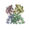
| ||||||||
|---|---|---|---|---|---|---|---|---|---|
| 1 |
| ||||||||
| Unit cell |
| ||||||||
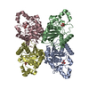
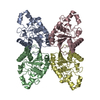
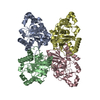
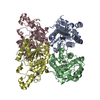
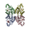
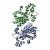
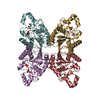
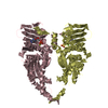
| Details | The biological assembly is the homotetramer formed by the chains A, B, C and D which forms the asymmetric unit. |
-Components
| #1: Protein | Mass: 28999.678 Da / Num. of mol.: 4 Source method: isolated from a genetically manipulated source Source: (gene. exp.) Mycobacterium tuberculosis (bacteria)Gene: GPMA, GPM, PGM, GPM1, RV0489, MT0508, MTCY20G9.15, MB0499 Plasmid: pET22b / Production host: References: UniProt: P0A5R6, UniProt: P9WIC9*PLUS, EC: 5.4.2.1 #2: Chemical | ChemComp-GOL /   Mass: 92.094 Da / Num. of mol.: 5 / Source method: obtained synthetically / Formula: C3H8O3 Mass: 92.094 Da / Num. of mol.: 5 / Source method: obtained synthetically / Formula: C3H8O3#3: Water | ChemComp-HOH / |  Mass: 18.015 Da / Num. of mol.: 451 / Source method: isolated from a natural source / Formula: H2O Mass: 18.015 Da / Num. of mol.: 451 / Source method: isolated from a natural source / Formula: H2O |
|---|
-Experimental details
-Experiment
| Experiment | Method: X-RAY DIFFRACTION / Number of used crystals: 1 |
|---|
- Sample preparation
| Crystal | Density Matthews: 2.51 Å3/Da / Density % sol: 45.79 % |
|---|---|
| Crystal grow | Temperature: 298 K / pH: 7 Details: 15% PEG 3350, 0.1M magnesium chloride, pH 7, VAPOR DIFFUSION, HANGING DROP, temperature 298K, pH 7.00 |
-Data collection
| Diffraction | Mean temperature: 100 K |
|---|---|
| Diffraction source | Source: SYNCHROTRON / Site: ALS  / Beamline: 8.2.2 / Wavelength: 1 / Beamline: 8.2.2 / Wavelength: 1 |
| Detector | Type: ADSC QUANTUM 315 / Detector: CCD / Date: Jul 11, 2003 |
| Radiation | Monochromator: DOUBLE CRYSTAL SI 111 / Protocol: SINGLE WAVELENGTH / Monochromatic (M) / Laue (L): M / Scattering type: x-ray |
| Radiation wavelength | Wavelength: 1 Å / Relative weight: 1 |
| Reflection | Resolution: 1.7→58.37 Å / Num. obs: 111293 / % possible obs: 98.2 % / Observed criterion σ(I): 0 / Redundancy: 6.5 % / Biso Wilson estimate: 21.33 Å2 / Rsym value: 0.0699 / Net I/σ(I): 18.1 |
| Reflection shell | Resolution: 1.7→1.8 Å / Redundancy: 3.7 % / Mean I/σ(I) obs: 3.2 / Rsym value: 0.4548 / % possible all: 91.8 |
- Processing
| Software |
| |||||||||||||||||||||||||||||||||
|---|---|---|---|---|---|---|---|---|---|---|---|---|---|---|---|---|---|---|---|---|---|---|---|---|---|---|---|---|---|---|---|---|---|---|
| Refinement | Method to determine structure: MOLECULAR REPLACEMENT Starting model: PDB ENTRY 5PGM Resolution: 1.7→58.37 Å / Num. parameters: 31673 / Num. restraintsaints: 31045 / Cross valid method: FREE R / σ(F): 0 / Stereochemistry target values: ENGH & HUBER Details: ANISOTROPIC SCALING APPLIED BY THE METHOD OF PARKIN, MOEZZI & HOPE, J.APPL.CRYST.28(1995)53-56. HYDROGENS HAVE BEEN ADDED AT GEOMETRICALLY CALCULATED POSITIONS AND REFINED USING A RIDING MODEL.
| |||||||||||||||||||||||||||||||||
| Solvent computation | Solvent model: Bulk solvent was treated following the Babinet-Principle refining two parameters, using the SWAT command in SHELXL. | |||||||||||||||||||||||||||||||||
| Displacement parameters | Biso mean: 33.8 Å2 | |||||||||||||||||||||||||||||||||
| Refine analyze | Num. disordered residues: 11 / Occupancy sum hydrogen: 6617 / Occupancy sum non hydrogen: 7846 | |||||||||||||||||||||||||||||||||
| Refinement step | Cycle: LAST / Resolution: 1.7→58.37 Å
| |||||||||||||||||||||||||||||||||
| Refine LS restraints |
| |||||||||||||||||||||||||||||||||
| LS refinement shell | Resolution: 1.7→1.8 Å /
|
