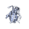
 Movie
Movie Controller


+ Open data

- Basic information
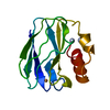
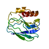
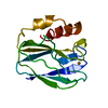
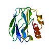
| Entry | Database: PDB / ID: 5paz | ||||||
|---|---|---|---|---|---|---|---|
| Title | REDUCED MUTANT P80A PSEUDOAZURIN FROM A. FAECALIS | ||||||
 Components Components | PSEUDOAZURIN | ||||||
 Keywords Keywords | ELECTRON TRANSFER / CUPROPROTEIN | ||||||
| Function / homology |  Function and homology information Function and homology information | ||||||
| Biological species |  Alcaligenes faecalis (bacteria) Alcaligenes faecalis (bacteria) | ||||||
| Method |  X-RAY DIFFRACTION / Resolution: 1.76 Å X-RAY DIFFRACTION / Resolution: 1.76 Å | ||||||
 Authors Authors | Adman, E.T. / Libeu, C.A.P. | ||||||
 Citation Citation | Journal: Biochemistry / Year: 1997 Title: Site-directed mutants of pseudoazurin: explanation of increased redox potentials from X-ray structures and from calculation of redox potential differences. Authors: Libeu, C.A. / Kukimoto, M. / Nishiyama, M. / Horinouchi, S. / Adman, E.T. #1: Journal: Protein Eng. / Year: 1992Title: Site-Directed Mutagenesis of Pseudoazurin from Alcaligenes Faecalis S-6; Pro80Ala Mutant Exhibits Marked Increase in Reduction Potential Authors: Nishiyama, M. / Suzuki, J. / Ohnuki, T. / Chang, H.C. / Horinouchi, S. / Turley, S. / Adman, E.T. / Beppu, T. #2: Journal: J.Biol.Chem. / Year: 1989Title: A 2.0-A Structure of the Blue Copper Protein (Cupredoxin) from Alcaligenes Faecalis S-6 Authors: Adman, E.T. / Turley, S. / Bramson, R. / Petratos, K. / Banner, D. / Tsernoglou, D. / Beppu, T. / Watanabe, H. | ||||||
| History |
|
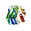
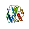
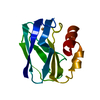
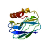
- Structure visualization
| Structure viewer | Molecule: MolmilJmol/JSmol |
|---|
- Downloads & links
-Download
| PDBx/mmCIF format | 5paz.cif.gz | 37.9 KB | Display | PDBx/mmCIF format |
|---|---|---|---|---|
| PDB format | pdb5paz.ent.gz | 25.5 KB | Display | PDB format |
| PDBx/mmJSON format | 5paz.json.gz | Tree view | PDBx/mmJSON format | |
| Others |  Other downloads Other downloads |
-Validation report
| Summary document | 5paz_validation.pdf.gz | 412.9 KB | Display | wwPDB validaton report |
|---|---|---|---|---|
| Full document | 5paz_full_validation.pdf.gz | 414 KB | Display | |
| Data in XML | 5paz_validation.xml.gz | 7.8 KB | Display | |
| Data in CIF | 5paz_validation.cif.gz | 10.1 KB | Display | |
| Arichive directory | https://data.pdbj.org/pub/pdb/validation_reports/pa/5pazftp://data.pdbj.org/pub/pdb/validation_reports/pa/5paz | HTTPS FTP |
-Related structure data


-Links
 PDBj
PDBj- Assembly
| Deposited unit | 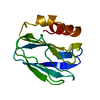
| ||||||||
|---|---|---|---|---|---|---|---|---|---|
| 1 |
| ||||||||
| Unit cell |
|
-Components
| #1: Protein | Mass: 13357.474 Da / Num. of mol.: 1 / Mutation: P80A Source method: isolated from a genetically manipulated source Source: (gene. exp.) Alcaligenes faecalis (bacteria) / Strain: S-6 / Cellular location: PERIPLASM / Gene: POTENTIAL / Production host: |
|---|---|
| #2: Chemical | ChemComp-CU /   Mass: 63.546 Da / Num. of mol.: 1 / Source method: obtained synthetically / Formula: Cu Mass: 63.546 Da / Num. of mol.: 1 / Source method: obtained synthetically / Formula: Cu |
| #3: Water | ChemComp-HOH /  Mass: 18.015 Da / Num. of mol.: 79 / Source method: isolated from a natural source / Formula: H2O Mass: 18.015 Da / Num. of mol.: 79 / Source method: isolated from a natural source / Formula: H2O |
-Experimental details
-Experiment
| Experiment | Method: X-RAY DIFFRACTION / Number of used crystals: 2 |
|---|
- Sample preparation
| Crystal | Density Matthews: 2.6 Å3/Da / Density % sol: 50 % | ||||||||||||||||||||
|---|---|---|---|---|---|---|---|---|---|---|---|---|---|---|---|---|---|---|---|---|---|
| Crystal grow | pH: 7 Details: 50MM SODIUM PHOSPHATE WITH 75% SATURATED AMMONIUM SULFATE, pH 7.0 | ||||||||||||||||||||
| Crystal grow | *PLUS pH: 6.8 / Method: unknown | ||||||||||||||||||||
| Components of the solutions | *PLUS
|
-Data collection
| Diffraction | Mean temperature: 298 K |
|---|---|
| Diffraction source | Source: ROTATING ANODE / Type: RIGAKU RUH2R / Wavelength: 1.5418 |
| Detector | Type: SIEMENS / Detector: AREA DETECTOR / Date: Jan 1, 1994 |
| Radiation | Monochromator: GRAPHITE(002) / Monochromatic (M) / Laue (L): M / Scattering type: x-ray |
| Radiation wavelength | Wavelength: 1.5418 Å / Relative weight: 1 |
| Reflection | Resolution: 1.76→10.3 Å / Num. obs: 10723 / Observed criterion σ(I): 0 / Rmerge(I) obs: 0.046 / Net I/σ(I): 21.5 |
| Reflection shell | Resolution: 1.76→1.8 Å / Rmerge(I) obs: 0.11 / Mean I/σ(I) obs: 8.1 |
| Reflection | *PLUS % possible obs: 84 % |
| Reflection shell | *PLUS % possible obs: 48 % |
- Processing
| Software |
| |||||||||||||||||||||||||||||||||
|---|---|---|---|---|---|---|---|---|---|---|---|---|---|---|---|---|---|---|---|---|---|---|---|---|---|---|---|---|---|---|---|---|---|---|
| Refinement | Resolution: 1.76→10.3 Å / Num. parameters: 4077 / Num. restraintsaints: 3791 / σ(F): 0 / Stereochemistry target values: ENGH AND HUBER Details: COPPER-LIGAND DISTANCES NOT RESTRAINED; CU + S REFINED ANISOTROPICALLY
| |||||||||||||||||||||||||||||||||
| Solvent computation | Solvent model: BABINET'S PRINCIPLE (SHELX-93) | |||||||||||||||||||||||||||||||||
| Refine analyze | Num. disordered residues: 0 | |||||||||||||||||||||||||||||||||
| Refinement step | Cycle: LAST / Resolution: 1.76→10.3 Å
| |||||||||||||||||||||||||||||||||
| Refine LS restraints |
| |||||||||||||||||||||||||||||||||
| Software | *PLUS Name: SHELXL / Classification: refinement | |||||||||||||||||||||||||||||||||
| Refinement | *PLUS Rfactor all: 0.17 / Rfactor obs: 0.163 | |||||||||||||||||||||||||||||||||
| Solvent computation | *PLUS | |||||||||||||||||||||||||||||||||
| Displacement parameters | *PLUS |
