 Movie
Movie Controller


+ Open data

- Basic information
| Entry | Database: PDB / ID: 1pzc | ||||||
|---|---|---|---|---|---|---|---|
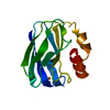
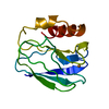
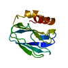
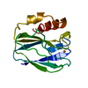
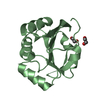
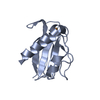
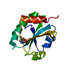
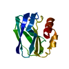
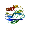
| Title | APO-PSEUDOAZURIN (METAL FREE PROTEIN) | ||||||
 Components Components | PSEUDOAZURIN | ||||||
 Keywords Keywords | ELECTRON TRANSFER (CUPROPROTEIN) / APOPROTEIN / METAL FREE PROTEIN | ||||||
| Function / homology |  Function and homology information Function and homology information | ||||||
| Biological species |  Alcaligenes faecalis (bacteria) Alcaligenes faecalis (bacteria) | ||||||
| Method |  X-RAY DIFFRACTION / SYNCHROTRON / Resolution: 1.85 Å X-RAY DIFFRACTION / SYNCHROTRON / Resolution: 1.85 Å | ||||||
 Authors Authors | Petratos, K. | ||||||
 Citation Citation | Journal: Febs Lett. / Year: 1995 Title: The crystal structure of apo-pseudoazurin from Alcaligenes faecalis S-6. Authors: Petratos, K. / Papadovasilaki, M. / Dauter, Z. #1: Journal: Acta Crystallogr.,Sect.B / Year: 1988Title: Refinement of the Structure of Pseudoazurin from Alcaligenes Faecalis S-6 at 1.55 Angstroms Resolution Authors: Petratos, K. / Dauter, Z. / Wilson, K.S. | ||||||
| History |
|
- Structure visualization
| Structure viewer | Molecule: MolmilJmol/JSmol |
|---|
- Downloads & links
-Download
| PDBx/mmCIF format | 1pzc.cif.gz | 37.7 KB | Display | PDBx/mmCIF format |
|---|---|---|---|---|
| PDB format | pdb1pzc.ent.gz | 26.1 KB | Display | PDB format |
| PDBx/mmJSON format | 1pzc.json.gz | Tree view | PDBx/mmJSON format | |
| Others |  Other downloads Other downloads |
-Validation report
| Arichive directory | https://data.pdbj.org/pub/pdb/validation_reports/pz/1pzcftp://data.pdbj.org/pub/pdb/validation_reports/pz/1pzc | HTTPS FTP |
|---|
-Related structure data
| Similar structure data |
|---|

-Links
 PDBj
PDBj- Assembly
| Deposited unit | 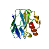
| ||||||||
|---|---|---|---|---|---|---|---|---|---|
| 1 |
| ||||||||
| Unit cell |
| ||||||||
| Atom site foot note | 1: CIS PROLINE - PRO 20 / 2: ATOM O HOH 136 WAS REFINED WITH OCCUPANCY SET TO 0.50. / 3: ATOM O HOH 137 WAS REFINED WITH OCCUPANCY SET TO 0.50. |
-Components
| #1: Protein | Mass: 13383.511 Da / Num. of mol.: 1 Source method: isolated from a genetically manipulated source Details: APO FORM / Source: (gene. exp.) Alcaligenes faecalis (bacteria) / Strain: S-6 / Plasmid: PUB1 / Production host: | ||
|---|---|---|---|
| #2: Water | ChemComp-HOH /  Mass: 18.015 Da / Num. of mol.: 137 / Source method: isolated from a natural source / Formula: H2O Mass: 18.015 Da / Num. of mol.: 137 / Source method: isolated from a natural source / Formula: H2O | ||
| Compound details | DE-METALLIZED| Source details | MOLECULE_NAME: PSEUDOAZURIN. THE PSEUDOAZURIN GENE FROM A. FAECALIS (438 B.P.) WAS EXPRESSED UNDER ...MOLECULE_NAME: PSEUDOAZUR | |
-Experimental details
-Experiment
| Experiment | Method: X-RAY DIFFRACTION / Number of used crystals: 1 |
|---|
- Sample preparation
| Crystal | Density Matthews: 2.61 Å3/Da / Density % sol: 52.95 % | ||||||||||||||||||||||||||||||
|---|---|---|---|---|---|---|---|---|---|---|---|---|---|---|---|---|---|---|---|---|---|---|---|---|---|---|---|---|---|---|---|
| Crystal grow | pH: 6.7 / Details: pH 6.7 | ||||||||||||||||||||||||||||||
| Crystal grow | *PLUS pH: 5.7 / Method: vapor diffusion / Details: Petratos, K., (1987) FEBS Lett., 218, 209. | ||||||||||||||||||||||||||||||
| Components of the solutions | *PLUS
|
-Data collection
| Diffraction source | Source: SYNCHROTRON / Site: EMBL/DESY, HAMBURG  / Beamline: BW7B / Wavelength: 0.87 Å / Beamline: BW7B / Wavelength: 0.87 Å |
|---|---|
| Detector | Type: MARRESEARCH / Detector: IMAGE PLATE / Date: Jul 13, 1994 |
| Radiation | Scattering type: x-ray |
| Radiation wavelength | Wavelength: 0.87 Å / Relative weight: 1 |
| Reflection | Num. obs: 11533 / % possible obs: 98.4 % / Observed criterion σ(I): 1 / Redundancy: 6.4 % / Rmerge(I) obs: 0.077 |
| Reflection | *PLUS Highest resolution: 1.85 Å / Lowest resolution: 10 Å / Num. obs: 11516 / Num. measured all: 73694 / Rmerge(I) obs: 0.077 |
- Processing
| Software |
| ||||||||||||||||||||||||||||||||||||||||||||||||||||||||||||||||||||||||||||||||
|---|---|---|---|---|---|---|---|---|---|---|---|---|---|---|---|---|---|---|---|---|---|---|---|---|---|---|---|---|---|---|---|---|---|---|---|---|---|---|---|---|---|---|---|---|---|---|---|---|---|---|---|---|---|---|---|---|---|---|---|---|---|---|---|---|---|---|---|---|---|---|---|---|---|---|---|---|---|---|---|---|---|
| Refinement | Resolution: 1.85→10 Å / σ(F): 1 /
| ||||||||||||||||||||||||||||||||||||||||||||||||||||||||||||||||||||||||||||||||
| Displacement parameters | Biso mean: 29 Å2 | ||||||||||||||||||||||||||||||||||||||||||||||||||||||||||||||||||||||||||||||||
| Refinement step | Cycle: LAST / Resolution: 1.85→10 Å
| ||||||||||||||||||||||||||||||||||||||||||||||||||||||||||||||||||||||||||||||||
| Refine LS restraints |
| ||||||||||||||||||||||||||||||||||||||||||||||||||||||||||||||||||||||||||||||||
| Refinement | *PLUS % reflection Rfree: 10 % | ||||||||||||||||||||||||||||||||||||||||||||||||||||||||||||||||||||||||||||||||
| Solvent computation | *PLUS | ||||||||||||||||||||||||||||||||||||||||||||||||||||||||||||||||||||||||||||||||
| Displacement parameters | *PLUS | ||||||||||||||||||||||||||||||||||||||||||||||||||||||||||||||||||||||||||||||||
| Refine LS restraints | *PLUS
|
