 Movie
Movie Controller


+ Open data

- Basic information
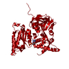
| Entry | Database: PDB / ID: 5oyi | ||||||
|---|---|---|---|---|---|---|---|
| Title | FMDV A10 dissociated pentamer | ||||||
 Components Components | (Genome polyprotein) x 3 | ||||||
 Keywords Keywords | VIRAL PROTEIN / A10 / FMDV / Pentamer / CryoEM / virus | ||||||
| Function / homology |  Function and homology information Function and homology informationL-peptidase / symbiont-mediated perturbation of host chromatin organization / picornain 3C / T=pseudo3 icosahedral viral capsid / host cell cytoplasmic vesicle membrane / ribonucleoside triphosphate phosphatase activity / nucleoside-triphosphate phosphatase / channel activity / monoatomic ion transmembrane transport / clathrin-dependent endocytosis of virus by host cell ...L-peptidase / symbiont-mediated perturbation of host chromatin organization / picornain 3C / T=pseudo3 icosahedral viral capsid / host cell cytoplasmic vesicle membrane / ribonucleoside triphosphate phosphatase activity / nucleoside-triphosphate phosphatase / channel activity / monoatomic ion transmembrane transport / clathrin-dependent endocytosis of virus by host cell / RNA helicase activity / viral protein processing / host cell endoplasmic reticulum membrane / symbiont-mediated activation of host autophagy / RNA-directed RNA polymerase / cysteine-type endopeptidase activity / viral RNA genome replication / RNA-directed RNA polymerase activity / DNA-templated transcription / virion attachment to host cell / host cell nucleus / structural molecule activity / proteolysis / RNA binding / ATP binding Similarity search - Function | ||||||
| Biological species |  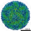 Foot-and-mouth disease virus Foot-and-mouth disease virus | ||||||
| Method | ELECTRON MICROSCOPY / single particle reconstruction / cryo EM / Resolution: 8.2 Å | ||||||
 Authors Authors | Kotecha, A. / Malik, N. / Stuart, D.I. | ||||||
| Funding support |  United Kingdom, 1items United Kingdom, 1items
| ||||||
 Citation Citation | Journal: PLoS Pathog / Year: 2017 Title: Structures of foot and mouth disease virus pentamers: Insight into capsid dissociation and unexpected pentamer reassociation. Authors: Nayab Malik / Abhay Kotecha / Sarah Gold / Amin Asfor / Jingshan Ren / Juha T Huiskonen / Tobias J Tuthill / Elizabeth E Fry / David I Stuart /  Abstract: Foot-and-mouth disease virus (FMDV) belongs to the Aphthovirus genus of the Picornaviridae, a family of small, icosahedral, non-enveloped, single-stranded RNA viruses. It is a highly infectious ...Foot-and-mouth disease virus (FMDV) belongs to the Aphthovirus genus of the Picornaviridae, a family of small, icosahedral, non-enveloped, single-stranded RNA viruses. It is a highly infectious pathogen and is one of the biggest hindrances to the international trade of animals and animal products. FMDV capsids (which are unstable below pH6.5) release their genome into the host cell from an acidic compartment, such as that of an endosome, and in the process dissociate into pentamers. Whilst other members of the family (enteroviruses) have been visualized to form an expanded intermediate capsid with holes from which inner capsid proteins (VP4), N-termini (VP1) and RNA can be released, there has been no visualization of any such state for an aphthovirus, instead the capsid appears to simply dissociate into pentamers. Here we present the 8-Å resolution structure of isolated dissociated pentamers of FMDV, lacking VP4. We also found these pentamers to re-associate into a rigid, icosahedrally symmetric assembly, which enabled their structure to be solved at higher resolution (5.2 Å). In this assembly, the pentamers unexpectedly associate 'inside out', but still with their exposed hydrophobic edges buried. Stabilizing interactions occur between the HI loop of VP2 and its symmetry related partners at the icosahedral 3-fold axes, and between the BC and EF loops of VP3 with the VP2 βB-strand and the CD loop at the 2-fold axes. A relatively extensive but subtle structural rearrangement towards the periphery of the dissociated pentamer compared to that in the mature virus provides insight into the mechanism of dissociation of FMDV and the marked difference in antigenicity. | ||||||
| History |
|
- Structure visualization
| Movie |
Movie viewer |
|---|---|
| Structure viewer | Molecule: MolmilJmol/JSmol |
- Downloads & links
-Download
| PDBx/mmCIF format | 5oyi.cif.gz | 554.7 KB | Display | PDBx/mmCIF format |
|---|---|---|---|---|
| PDB format | pdb5oyi.ent.gz | 454.8 KB | Display | PDB format |
| PDBx/mmJSON format | 5oyi.json.gz | Tree view | PDBx/mmJSON format | |
| Others |  Other downloads Other downloads |
-Validation report
| Arichive directory | https://data.pdbj.org/pub/pdb/validation_reports/oy/5oyiftp://data.pdbj.org/pub/pdb/validation_reports/oy/5oyi | HTTPS FTP |
|---|
-Related structure data
| Related structure data | 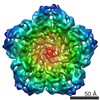 3862MC 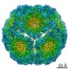 3856C  5owxC C: citing same article ( M: map data used to model this data |
|---|---|
| Similar structure data |

-Links
 PDBj
PDBj

- Assembly
| Deposited unit | 
|
|---|---|
| 1 |
|
-Components
| #1: Protein | Mass: 20097.980 Da / Num. of mol.: 5 Source method: isolated from a genetically manipulated source Source: (gene. exp.) Foot-and-mouth disease virus (strain A10-61)Cell line (production host): BHK21 / Production host:  Cricetinae gen. sp. (mammal) Cricetinae gen. sp. (mammal)References: UniProt: P03306, L-peptidase, nucleoside-triphosphate phosphatase, picornain 3C, RNA-directed RNA polymerase #2: Protein | Mass: 20701.512 Da / Num. of mol.: 5 Source method: isolated from a genetically manipulated source Source: (gene. exp.) Foot-and-mouth disease virus (strain A10-61)Production host: Cricetinae gen. sp. (mammal)References: UniProt: P03306, L-peptidase, nucleoside-triphosphate phosphatase, picornain 3C, RNA-directed RNA polymerase #3: Protein | Mass: 24233.992 Da / Num. of mol.: 5 Source method: isolated from a genetically manipulated source Source: (gene. exp.) Foot-and-mouth disease virus (strain A10-61)Production host: Cricetinae gen. sp. (mammal)References: UniProt: P03306, L-peptidase, nucleoside-triphosphate phosphatase, picornain 3C, RNA-directed RNA polymerase |
|---|
-Experimental details
-Experiment
| Experiment | Method: ELECTRON MICROSCOPY |
|---|---|
| EM experiment | Aggregation state: PARTICLE / 3D reconstruction method: single particle reconstruction |
- Sample preparation
| Component | Name: FMDV A10 dissociated pentamer / Type: VIRUS / Entity ID: all / Source: RECOMBINANT |
|---|---|
| Molecular weight | Value: 455 kDa/nm / Experimental value: NO |
| Source (natural) | Organism: Foot-and-mouth disease virus (strain A10-61) |
| Source (recombinant) | Organism: Cricetinae gen. sp. (mammal) |
| Details of virus | Empty: NO / Enveloped: NO / Isolate: SEROTYPE / Type: VIRION |
| Natural host | Organism: Bos taurus |
| Virus shell | Name: Foot and Disease Virus A1061 / Diameter: 30 nm / Triangulation number (T number): 3 |
| Buffer solution | pH: 8 / Details: 50mM Hepes, 200mM NaCl |
| Specimen | Conc.: 2.5 mg/ml / Embedding applied: NO / Shadowing applied: NO / Staining applied: NO / Vitrification applied: YES |
| Specimen support | Grid material: COPPER / Grid mesh size: 200 divisions/in. / Grid type: C-Flats R2/1 |
| Vitrification | Instrument: FEI VITROBOT MARK IV / Cryogen name: ETHANE / Humidity: 90 % / Chamber temperature: 298 K |
- Electron microscopy imaging
| Experimental equipment |  Model: Tecnai Polara / Image courtesy: FEI Company |
|---|---|
| Microscopy | Model: FEI POLARA 300 |
| Electron gun | Electron source:  FIELD EMISSION GUN / Accelerating voltage: 300 kV / Illumination mode: FLOOD BEAM FIELD EMISSION GUN / Accelerating voltage: 300 kV / Illumination mode: FLOOD BEAM |
| Electron lens | Mode: BRIGHT FIELD / Nominal magnification: 160000 X / Calibrated magnification: 37037 X / Nominal defocus max: 5000 nm / Nominal defocus min: 1200 nm / Cs: 2.7 mm / C2 aperture diameter: 50 µm / Alignment procedure: BASIC |
| Specimen holder | Cryogen: NITROGEN Specimen holder model: GATAN 910 MULTI-SPECIMEN SINGLE TILT CRYO TRANSFER HOLDER Temperature (max): 70 K / Temperature (min): 70 K |
| Image recording | Average exposure time: 5 sec. / Electron dose: 20 e/Å2 / Detector mode: COUNTING / Film or detector model: GATAN K2 SUMMIT (4k x 4k) / Num. of grids imaged: 1 / Num. of real images: 1570 |
| EM imaging optics | Energyfilter name: GIF / Energyfilter upper: 20 eV / Energyfilter lower: 0 eV |
| Image scans | Sampling size: 5 µm / Width: 3838 / Height: 3710 / Movie frames/image: 25 / Used frames/image: 2-20 |
- Processing
| EM software |
| ||||||||||||||||||||||||||||||||||||||||
|---|---|---|---|---|---|---|---|---|---|---|---|---|---|---|---|---|---|---|---|---|---|---|---|---|---|---|---|---|---|---|---|---|---|---|---|---|---|---|---|---|---|
| CTF correction | Type: PHASE FLIPPING AND AMPLITUDE CORRECTION | ||||||||||||||||||||||||||||||||||||||||
| Symmetry | Point symmetry: C5 (5 fold cyclic) | ||||||||||||||||||||||||||||||||||||||||
| 3D reconstruction | Resolution: 8.2 Å / Resolution method: FSC 0.143 CUT-OFF / Num. of particles: 4260 / Algorithm: BACK PROJECTION / Num. of class averages: 1 / Symmetry type: POINT | ||||||||||||||||||||||||||||||||||||||||
| Atomic model building | B value: 200 / Protocol: RIGID BODY FIT / Space: REAL / Target criteria: Cross-correlation coefficient |
