 Movie
Movie Controller

[English] 日本語
 Yorodumi
- PDB-5oxs: Crystal structure of human lung surfactant protein D trimeric fra... -
+ Open data

- Basic information
| Entry | Database: PDB / ID: 5oxs | |||||||||
|---|---|---|---|---|---|---|---|---|---|---|
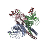
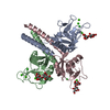
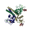
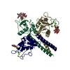
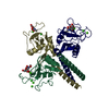
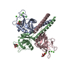
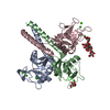
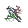
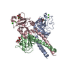
| Title | Crystal structure of human lung surfactant protein D trimeric fragment with bound ligand Salmonella enterica Minnesota R5 oligosaccharide | |||||||||
 Components Components | Pulmonary surfactant-associated protein D | |||||||||
 Keywords Keywords | SURFACTANT PROTEIN / trimeric recombinant collectin fragment / neck+CRD / alpha-helical coiled coil / carbohydrate recognition domain / lectin / sugar binding protein | |||||||||
| Function / homology |  Function and homology information Function and homology informationToll Like Receptor TLR1:TLR2 Cascade / Defective CSF2RB causes SMDP5 / Defective CSF2RA causes SMDP4 / Toll Like Receptor 4 (TLR4) Cascade / clathrin-coated endocytic vesicle / respiratory gaseous exchange by respiratory system / collagen trimer / Regulation of TLR by endogenous ligand / surfactant homeostasis / Surfactant metabolism ...Toll Like Receptor TLR1:TLR2 Cascade / Defective CSF2RB causes SMDP5 / Defective CSF2RA causes SMDP4 / Toll Like Receptor 4 (TLR4) Cascade / clathrin-coated endocytic vesicle / respiratory gaseous exchange by respiratory system / collagen trimer / Regulation of TLR by endogenous ligand / surfactant homeostasis / Surfactant metabolism / Signal regulatory protein family interactions / negative regulation of interleukin-2 production / lung alveolus development / macrophage chemotaxis / endocytic vesicle / negative regulation of T cell proliferation / multivesicular body / regulation of cytokine production / reactive oxygen species metabolic process / positive regulation of phagocytosis / receptor-mediated endocytosis / Immunoregulatory interactions between a Lymphoid and a non-Lymphoid cell / SARS-CoV-1 activates/modulates innate immune responses / carbohydrate binding / lysosome / defense response to bacterium / innate immune response / endoplasmic reticulum membrane / SARS-CoV-2 activates/modulates innate and adaptive immune responses / : / extracellular region / identical protein binding Similarity search - Function | |||||||||
| Biological species |  Homo sapiens (human) Homo sapiens (human) | |||||||||
| Method |  X-RAY DIFFRACTION / SYNCHROTRON / MOLECULAR REPLACEMENT / Resolution: 1.65 Å X-RAY DIFFRACTION / SYNCHROTRON / MOLECULAR REPLACEMENT / Resolution: 1.65 Å | |||||||||
 Authors Authors | Shrive, A.K. / Greenhough, T.J. | |||||||||
 Citation Citation | Journal: PLoS ONE / Year: 2018 Title: Structural definition of hSP-D recognition of Salmonella enterica LPS inner core oligosaccharides reveals alternative binding modes for the same LPS. Authors: Littlejohn, J.R. / da Silva, R.F. / Neale, W.A. / Smallcombe, C.C. / Clark, H.W. / Mackay, R.A. / Watson, A.S. / Madsen, J. / Hood, D.W. / Burns, I. / Greenhough, T.J. / Shrive, A.K. #1: Journal: Infect. Immun. / Year: 2016Title: Crystal Structure of a Complex of Surfactant Protein D (SP-D) and Haemophilus influenzae Lipopolysaccharide Reveals Shielding of Core Structures in SP-D-Resistant Strains. Authors: Clark, H.W. / Mackay, R.M. / Deadman, M.E. / Hood, D.W. / Madsen, J. / Moxon, E.R. / Townsend, J.P. / Reid, K.B.M. / Ahmed, A. / Shaw, A.J. / Greenhough, T.J. / Shrive, A.K. | |||||||||
| History |
|
- Structure visualization
| Structure viewer | Molecule: MolmilJmol/JSmol |
|---|
- Downloads & links
-Download
| PDBx/mmCIF format | 5oxs.cif.gz | 120.3 KB | Display | PDBx/mmCIF format |
|---|---|---|---|---|
| PDB format | pdb5oxs.ent.gz | 90.3 KB | Display | PDB format |
| PDBx/mmJSON format | 5oxs.json.gz | Tree view | PDBx/mmJSON format | |
| Others |  Other downloads Other downloads |
-Validation report
| Arichive directory | https://data.pdbj.org/pub/pdb/validation_reports/ox/5oxsftp://data.pdbj.org/pub/pdb/validation_reports/ox/5oxs | HTTPS FTP |
|---|
-Related structure data
| Related structure data |  5oxrC  1pw9S C: citing same article ( S: Starting model for refinement |
|---|---|
| Similar structure data |

-Links
 PDBj
PDBj


- Assembly
| Deposited unit | 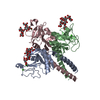
| ||||||||
|---|---|---|---|---|---|---|---|---|---|
| 1 |
| ||||||||
| Unit cell |
|
-Components
| #1: Protein | Mass: 18834.957 Da / Num. of mol.: 3 / Fragment: Trimeric neck + carbohydrate recognition domain Source method: isolated from a genetically manipulated source Source: (gene. exp.) Homo sapiens (human) / Gene: SFTPD, COLEC7, PSPD, SFTP4 / Production host:  #2: Polysaccharide | Source method: isolated from a genetically manipulated source #3: Chemical | ChemComp-CA /   Mass: 40.078 Da / Num. of mol.: 5 / Source method: obtained synthetically / Formula: Ca Mass: 40.078 Da / Num. of mol.: 5 / Source method: obtained synthetically / Formula: Ca#4: Water | ChemComp-HOH / |  Mass: 18.015 Da / Num. of mol.: 477 / Source method: isolated from a natural source / Formula: H2O Mass: 18.015 Da / Num. of mol.: 477 / Source method: isolated from a natural source / Formula: H2OHas protein modification | Y | |
|---|
-Experimental details
-Experiment
| Experiment | Method: X-RAY DIFFRACTION / Number of used crystals: 1 |
|---|
- Sample preparation
| Crystal | Density Matthews: 3 Å3/Da / Density % sol: 58.99 % / Mosaicity: 0.34 ° |
|---|---|
| Crystal grow | Temperature: 293 K / Method: vapor diffusion, sitting drop / pH: 8 / Details: 0.1M Tris pH 8.0, 16% PEG 6000 |
-Data collection
| Diffraction | Mean temperature: 100 K | ||||||||||||||||||||||||
|---|---|---|---|---|---|---|---|---|---|---|---|---|---|---|---|---|---|---|---|---|---|---|---|---|---|
| Diffraction source | Source: SYNCHROTRON / Site: Diamond  / Beamline: I04-1 / Wavelength: 0.92819 Å / Beamline: I04-1 / Wavelength: 0.92819 Å | ||||||||||||||||||||||||
| Detector | Type: DECTRIS PILATUS 6M-F / Detector: PIXEL / Date: May 16, 2016 | ||||||||||||||||||||||||
| Radiation | Protocol: SINGLE WAVELENGTH / Monochromatic (M) / Laue (L): M / Scattering type: x-ray | ||||||||||||||||||||||||
| Radiation wavelength | Wavelength: 0.92819 Å / Relative weight: 1 | ||||||||||||||||||||||||
| Reflection | Resolution: 1.65→56.1 Å / Num. obs: 74199 / % possible obs: 93 % / Redundancy: 3.1 % / CC1/2: 0.998 / Rmerge(I) obs: 0.054 / Rpim(I) all: 0.036 / Rrim(I) all: 0.065 / Net I/σ(I): 11.4 | ||||||||||||||||||||||||
| Reflection shell | Diffraction-ID: 1
|
- Processing
| Software |
| ||||||||||||||||||||||||||||||||||||||||||||||||||||||||||||
|---|---|---|---|---|---|---|---|---|---|---|---|---|---|---|---|---|---|---|---|---|---|---|---|---|---|---|---|---|---|---|---|---|---|---|---|---|---|---|---|---|---|---|---|---|---|---|---|---|---|---|---|---|---|---|---|---|---|---|---|---|---|
| Refinement | Method to determine structure: MOLECULAR REPLACEMENT Starting model: pdbid 1PW9 Resolution: 1.65→56.1 Å / Cor.coef. Fo:Fc: 0.969 / Cor.coef. Fo:Fc free: 0.961 / SU B: 1.683 / SU ML: 0.055 / SU R Cruickshank DPI: 0.0789 / Cross valid method: THROUGHOUT / σ(F): 0 / ESU R: 0.079 / ESU R Free: 0.078 Details: HYDROGENS HAVE BEEN ADDED IN THE RIDING POSITIONS U VALUES : REFINED INDIVIDUALLY
| ||||||||||||||||||||||||||||||||||||||||||||||||||||||||||||
| Solvent computation | Ion probe radii: 0.8 Å / Shrinkage radii: 0.8 Å / VDW probe radii: 1.2 Å | ||||||||||||||||||||||||||||||||||||||||||||||||||||||||||||
| Displacement parameters | Biso max: 98.23 Å2 / Biso mean: 23.384 Å2 / Biso min: 11.59 Å2
| ||||||||||||||||||||||||||||||||||||||||||||||||||||||||||||
| Refinement step | Cycle: final / Resolution: 1.65→56.1 Å
| ||||||||||||||||||||||||||||||||||||||||||||||||||||||||||||
| Refine LS restraints |
| ||||||||||||||||||||||||||||||||||||||||||||||||||||||||||||
| LS refinement shell | Resolution: 1.65→1.693 Å / Rfactor Rfree error: 0 / Total num. of bins used: 20
|
