 Movie
Movie Controller

[English] 日本語
 Yorodumi
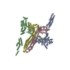
- PDB-5oxf: An oligomerised bacterial dynamin pair provides a mechanism for t... -
+ Open data

- Basic information
| Entry | Database: PDB / ID: 5oxf | ||||||
|---|---|---|---|---|---|---|---|
| Title | An oligomerised bacterial dynamin pair provides a mechanism for the long range sensing and tethering of membranes | ||||||
 Components Components | (GTP-binding protein) x 2 | ||||||
 Keywords Keywords | LIPID BINDING PROTEIN / dynamin / membrane remodelling / membrane fusion / membrane tethering. | ||||||
| Function / homology |  Function and homology information Function and homology information | ||||||
| Biological species |   Campylobacter jejuni (Campylobacter) Campylobacter jejuni (Campylobacter) | ||||||
| Method |  X-RAY DIFFRACTION / SYNCHROTRON / SAD / Resolution: 3.94 Å X-RAY DIFFRACTION / SYNCHROTRON / SAD / Resolution: 3.94 Å | ||||||
 Authors Authors | Liu, J.W. / Noel, J.K. / Low, H.H. | ||||||
| Funding support |  United Kingdom, 1items United Kingdom, 1items
| ||||||
 Citation Citation | Journal: Nat Commun / Year: 2018 Title: Structural basis for membrane tethering by a bacterial dynamin-like pair. Authors: Liu, J. / Noel, J.K. / Low, H.H. | ||||||
| History |
|
- Structure visualization
| Structure viewer | Molecule: MolmilJmol/JSmol |
|---|
- Downloads & links
-Download
| PDBx/mmCIF format | 5oxf.cif.gz | 510.1 KB | Display | PDBx/mmCIF format |
|---|---|---|---|---|
| PDB format | pdb5oxf.ent.gz | 407.4 KB | Display | PDB format |
| PDBx/mmJSON format | 5oxf.json.gz | Tree view | PDBx/mmJSON format | |
| Others |  Other downloads Other downloads |
-Validation report
| Arichive directory | https://data.pdbj.org/pub/pdb/validation_reports/ox/5oxfftp://data.pdbj.org/pub/pdb/validation_reports/ox/5oxf | HTTPS FTP |
|---|
-Related structure data

-Links
 PDBj
PDBj- Assembly
| Deposited unit | 
| ||||||||
|---|---|---|---|---|---|---|---|---|---|
| 1 |
| ||||||||
| Unit cell |
|
-Components
| #1: Protein | Mass: 85217.000 Da / Num. of mol.: 2 Source method: isolated from a genetically manipulated source Details: GDP / Source: (gene. exp.) Campylobacter jejuni (Campylobacter) / Gene: BKM79_02020, CRM98_01430 / Production host: #2: Protein | Mass: 71951.148 Da / Num. of mol.: 2 Source method: isolated from a genetically manipulated source Source: (gene. exp.) Campylobacter jejuni (Campylobacter) / Gene: BKM79_02025 / Production host: #3: Chemical | ChemComp-GDP /   Type: RNA linking / Mass: 443.201 Da / Num. of mol.: 4 / Source method: obtained synthetically / Formula: C10H15N5O11P2 / Comment: GDP, energy-carrying molecule*YM Type: RNA linking / Mass: 443.201 Da / Num. of mol.: 4 / Source method: obtained synthetically / Formula: C10H15N5O11P2 / Comment: GDP, energy-carrying molecule*YM |
|---|
-Experimental details
-Experiment
| Experiment | Method: X-RAY DIFFRACTION / Number of used crystals: 1 |
|---|
- Sample preparation
| Crystal | Density Matthews: 6.65 Å3/Da / Density % sol: 81.51 % |
|---|---|
| Crystal grow | Temperature: 293.15 K / Method: vapor diffusion, sitting drop / Details: 0.7-1.0 M succinate pH 7.0 |
-Data collection
| Diffraction | Mean temperature: 100 K |
|---|---|
| Diffraction source | Source: SYNCHROTRON / Site: Diamond / Beamline: I04-1 / Wavelength: 0.92819 Å |
| Detector | Type: DECTRIS PILATUS 6M-F / Detector: PIXEL / Date: Aug 8, 2015 |
| Radiation | Protocol: SINGLE WAVELENGTH / Monochromatic (M) / Laue (L): M / Scattering type: x-ray |
| Radiation wavelength | Wavelength: 0.92819 Å / Relative weight: 1 |
| Reflection | Resolution: 3.94→185.8 Å / Num. obs: 74962 / % possible obs: 95.5 % / Redundancy: 3.8 % / Net I/σ(I): 14.6 |
| Reflection shell | Resolution: 3.94→4.08 Å |
- Processing
| Software |
| ||||||||||||||||||||||||||||||||||||||||||||||||||||||||||||||||||||||||||||||||||||||||||||||||||||||||||||||||||||||||||||||||||||||||||||||||||||||||||||||||||||||||||||||||||||||
|---|---|---|---|---|---|---|---|---|---|---|---|---|---|---|---|---|---|---|---|---|---|---|---|---|---|---|---|---|---|---|---|---|---|---|---|---|---|---|---|---|---|---|---|---|---|---|---|---|---|---|---|---|---|---|---|---|---|---|---|---|---|---|---|---|---|---|---|---|---|---|---|---|---|---|---|---|---|---|---|---|---|---|---|---|---|---|---|---|---|---|---|---|---|---|---|---|---|---|---|---|---|---|---|---|---|---|---|---|---|---|---|---|---|---|---|---|---|---|---|---|---|---|---|---|---|---|---|---|---|---|---|---|---|---|---|---|---|---|---|---|---|---|---|---|---|---|---|---|---|---|---|---|---|---|---|---|---|---|---|---|---|---|---|---|---|---|---|---|---|---|---|---|---|---|---|---|---|---|---|---|---|---|---|
| Refinement | Method to determine structure: SAD / Resolution: 3.94→58.981 Å / SU ML: 0.5 / Cross valid method: FREE R-VALUE / σ(F): 1.34 / Phase error: 31.75 / Stereochemistry target values: ML
| ||||||||||||||||||||||||||||||||||||||||||||||||||||||||||||||||||||||||||||||||||||||||||||||||||||||||||||||||||||||||||||||||||||||||||||||||||||||||||||||||||||||||||||||||||||||
| Solvent computation | Shrinkage radii: 0.9 Å / VDW probe radii: 1.11 Å / Solvent model: FLAT BULK SOLVENT MODEL | ||||||||||||||||||||||||||||||||||||||||||||||||||||||||||||||||||||||||||||||||||||||||||||||||||||||||||||||||||||||||||||||||||||||||||||||||||||||||||||||||||||||||||||||||||||||
| Refinement step | Cycle: LAST / Resolution: 3.94→58.981 Å
| ||||||||||||||||||||||||||||||||||||||||||||||||||||||||||||||||||||||||||||||||||||||||||||||||||||||||||||||||||||||||||||||||||||||||||||||||||||||||||||||||||||||||||||||||||||||
| Refine LS restraints |
| ||||||||||||||||||||||||||||||||||||||||||||||||||||||||||||||||||||||||||||||||||||||||||||||||||||||||||||||||||||||||||||||||||||||||||||||||||||||||||||||||||||||||||||||||||||||
| LS refinement shell |
|
