| Entry | Database: PDB / ID: 5ox3
|
|---|
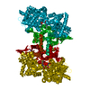
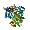
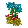
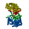
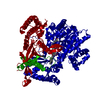
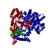
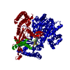
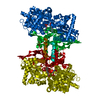
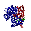
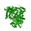
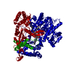
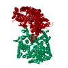
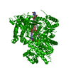
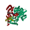
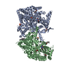
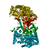
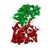
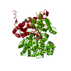
| Title | Glycogen Phosphorylase in complex with SzB102v |
|---|
 Components Components | Glycogen phosphorylase, muscle form |
|---|
 Keywords Keywords | TRANSFERASE |
|---|
| Function / homology |  Function and homology information Function and homology information
glycogen phosphorylase / glycogen phosphorylase activity / glycogen catabolic process / skeletal muscle myofibril / pyridoxal phosphate binding / nucleotide bindingSimilarity search - Function Glycogen/starch/alpha-glucan phosphorylase / Phosphorylase pyridoxal-phosphate attachment site / Phosphorylase pyridoxal-phosphate attachment site. / Glycosyl transferase, family 35 / Carbohydrate phosphorylase / Glycogen Phosphorylase B; / Rossmann fold / 3-Layer(aba) Sandwich / Alpha BetaSimilarity search - Domain/homology |
|---|
| Biological species |   Oryctolagus cuniculus (rabbit) Oryctolagus cuniculus (rabbit) |
|---|
| Method |  X-RAY DIFFRACTION / SYNCHROTRON / FOURIER SYNTHESIS / Resolution: 1.9 Å X-RAY DIFFRACTION / SYNCHROTRON / FOURIER SYNTHESIS / Resolution: 1.9 Å |
|---|
 Authors Authors | Kyriakis, E. / Stravodimos, G.A. / Kantsadi, A.L. / Chatzileontiadou, D.S.M. / Leonidas, D.D. |
|---|
 Citation Citation | Journal: Bioorg. Chem. / Year: 2018
Title: Probing the beta-pocket of the active site of human liver glycogen phosphorylase with 3-(C-beta-d-glucopyranosyl)-5-(4-substituted-phenyl)-1, 2, 4-triazole inhibitors.
Authors: Kyriakis, E. / Solovou, T.G.A. / Kun, S. / Czifrak, K. / Szocs, B. / Juhasz, L. / Bokor, E. / Stravodimos, G.A. / Kantsadi, A.L. / Chatzileontiadou, D.S.M. / Skamnaki, V.T. / Somsak, L. / Leonidas, D.D. |
|---|
| History | | Deposition | Sep 5, 2017 | Deposition site: PDBE / Processing site: PDBE |
|---|
| Revision 1.0 | Feb 28, 2018 | Provider: repository / Type: Initial release |
|---|
| Revision 2.0 | Jul 29, 2020 | Group: Atomic model / Derived calculations / Structure summary
Category: atom_site / chem_comp ...atom_site / chem_comp / entity / pdbx_entity_nonpoly / struct_site / struct_site_gen
Item: _atom_site.auth_atom_id / _atom_site.label_atom_id ..._atom_site.auth_atom_id / _atom_site.label_atom_id / _chem_comp.name / _chem_comp.pdbx_synonyms / _chem_comp.type / _entity.pdbx_description / _pdbx_entity_nonpoly.name
Description: Carbohydrate remediation / Provider: repository / Type: Remediation |
|---|
| Revision 2.1 | Apr 9, 2025 | Group: Data collection / Database references / Structure summary
Category: chem_comp_atom / chem_comp_bond ...chem_comp_atom / chem_comp_bond / database_2 / pdbx_entry_details
Item: _database_2.pdbx_DOI / _database_2.pdbx_database_accession |
|---|
|
|---|
 Movie
Movie Controller


 Open data
 Basic information
 Structure visualization
 Downloads & links
 Other downloads


 PDBj
PDBj
 Assembly

 Type: D-saccharide / Mass: 323.301 Da / Num. of mol.: 1 / Source method: obtained synthetically / Formula: C14H17N3O6
 Mass: 247.142 Da / Num. of mol.: 1 / Source method: obtained synthetically / Formula: C8H10NO6P
 Mass: 348.206 Da / Num. of mol.: 1 / Source method: obtained synthetically / Formula: C10H13N4O8P
 Mass: 18.015 Da / Num. of mol.: 268 / Source method: isolated from a natural source / Formula: H2O
 Sample preparation
 / Beamline: I911-2 / Wavelength: 1.0403 Å
 Processing