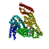
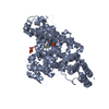
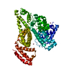
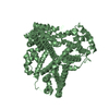
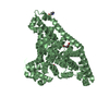
Entry Database : PDB / ID : 5oswTitle Structure of caprine serum albumin in complex with 3,5-diiodosalicylic acid Albumin Keywords / / / / / / Function / homology Function Domain/homology Component
/ / / / / / / / / / / / / / / / / / / / / Biological species Capra hircus (goat)Method / / / Resolution : 1.78 Å Authors Talaj, J.A. / Bujacz, A. / Bujacz, G. Funding support Organization Grant number Country National Science Centre 2013/11/B/ST5/02271
Journal : Acta Crystallogr D Struct Biol / Year : 2017Title : Crystal structures of serum albumins from domesticated ruminants and their complexes with 3,5-diiodosalicylic acid.Authors : Bujacz, A. / Talaj, J.A. / Zielinski, K. / Pietrzyk-Brzezinska, A.J. / Neumann, P. History Deposition Aug 18, 2017 Deposition site / Processing site Revision 1.0 Nov 8, 2017 Provider / Type Revision 1.1 Nov 22, 2017 Group / Category Item _citation.country / _citation.journal_abbrev ... _citation.country / _citation.journal_abbrev / _citation.journal_id_ASTM / _citation.journal_id_CSD / _citation.journal_id_ISSN / _citation.pdbx_database_id_PubMed / _citation.title Revision 1.2 Jan 17, 2024 Group / Database references / Refinement descriptionCategory chem_comp_atom / chem_comp_bond ... chem_comp_atom / chem_comp_bond / database_2 / pdbx_initial_refinement_model Item / _database_2.pdbx_database_accessionRevision 1.3 Nov 20, 2024 Group / Category / pdbx_modification_feature
Show all Show less
 Movie
Movie Controller

 Yorodumi
 Open data
 Basic information
 Components
 Keywords
 Function and homology information
 X-RAY DIFFRACTION /
  Authors
 Poland, 1items
  Citation
 Structure visualization
 Downloads & links
 Other downloads


 PDBj
PDBj


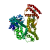
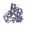
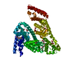
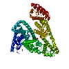
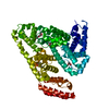
 Assembly
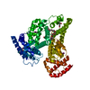


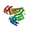
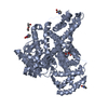
 Mass: 389.914 Da / Num. of mol.: 6 / Source method: obtained synthetically / Formula: C7H4I2O3
 Mass: 192.124 Da / Num. of mol.: 1 / Source method: obtained synthetically / Formula: C6H8O7
 Mass: 597.822 Da / Num. of mol.: 1 / Source method: obtained synthetically / Formula: C30H63NO10
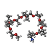
 Mass: 266.331 Da / Num. of mol.: 1 / Source method: obtained synthetically / Formula: C12H26O6
 Sample preparation
 / Beamline: 14.2 / Wavelength: 0.9184 Å
 Processing