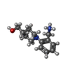
+ データを開く
- 基本情報
基本情報
| 登録情報 | データベース: PDB / ID: 5os2 | |||||||||
|---|---|---|---|---|---|---|---|---|---|---|
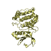
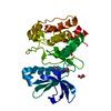
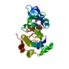
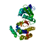
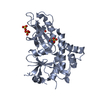
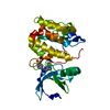
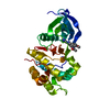
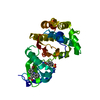
| タイトル | Crystal structure of Aurora-A kinase in complex with an allosterically binding fragment | |||||||||
 要素 要素 | Aurora kinase A | |||||||||
 キーワード キーワード | TRANSFERASE / kinase / allosteric inhibitor / fragment | |||||||||
| 機能・相同性 |  機能・相同性情報 機能・相同性情報Interaction between PHLDA1 and AURKA / regulation of centrosome cycle / axon hillock / spindle assembly involved in female meiosis I / cilium disassembly / spindle pole centrosome / chromosome passenger complex / histone H3S10 kinase activity / positive regulation of oocyte maturation / mitotic centrosome separation ...Interaction between PHLDA1 and AURKA / regulation of centrosome cycle / axon hillock / spindle assembly involved in female meiosis I / cilium disassembly / spindle pole centrosome / chromosome passenger complex / histone H3S10 kinase activity / positive regulation of oocyte maturation / mitotic centrosome separation / pronucleus / germinal vesicle / protein localization to centrosome / meiotic spindle / anterior/posterior axis specification / neuron projection extension / spindle organization / centrosome localization / positive regulation of mitochondrial fission / mitotic spindle pole / spindle midzone / SUMOylation of DNA replication proteins / negative regulation of protein binding / regulation of G2/M transition of mitotic cell cycle / protein serine/threonine/tyrosine kinase activity / liver regeneration / centriole / positive regulation of mitotic nuclear division / positive regulation of mitotic cell cycle / TP53 Regulates Transcription of Genes Involved in G2 Cell Cycle Arrest / molecular function activator activity / regulation of signal transduction by p53 class mediator / AURKA Activation by TPX2 / mitotic spindle organization / regulation of cytokinesis / APC/C:Cdh1 mediated degradation of Cdc20 and other APC/C:Cdh1 targeted proteins in late mitosis/early G1 / FBXL7 down-regulates AURKA during mitotic entry and in early mitosis / peptidyl-serine phosphorylation / regulation of protein stability / kinetochore / response to wounding / G2/M transition of mitotic cell cycle / spindle / spindle pole / mitotic spindle / Regulation of PLK1 Activity at G2/M Transition / positive regulation of proteasomal ubiquitin-dependent protein catabolic process / mitotic cell cycle / protein autophosphorylation / microtubule cytoskeleton / midbody / Regulation of TP53 Activity through Phosphorylation / basolateral plasma membrane / proteasome-mediated ubiquitin-dependent protein catabolic process / microtubule / protein phosphorylation / non-specific serine/threonine protein kinase / protein kinase activity / postsynaptic density / ciliary basal body / protein heterodimerization activity / negative regulation of gene expression / cell division / protein serine kinase activity / protein serine/threonine kinase activity / apoptotic process / ubiquitin protein ligase binding / centrosome / protein kinase binding / negative regulation of apoptotic process / perinuclear region of cytoplasm / glutamatergic synapse / nucleoplasm / ATP binding / nucleus / cytosol 類似検索 - 分子機能 | |||||||||
| 生物種 |  Homo sapiens (ヒト) Homo sapiens (ヒト) | |||||||||
| 手法 |  X線回折 / X線回折 /  シンクロトロン / 解像度: 1.92 Å シンクロトロン / 解像度: 1.92 Å | |||||||||
 データ登録者 データ登録者 | McIntyre, P.J. / Collins, P.M. / von Delft, F. / Bayliss, R. | |||||||||
| 資金援助 |  英国, 2件 英国, 2件
| |||||||||
 引用 引用 |  ジャーナル: ACS Chem. Biol. / 年: 2017 ジャーナル: ACS Chem. Biol. / 年: 2017タイトル: Characterization of Three Druggable Hot-Spots in the Aurora-A/TPX2 Interaction Using Biochemical, Biophysical, and Fragment-Based Approaches. 著者: McIntyre, P.J. / Collins, P.M. / Vrzal, L. / Birchall, K. / Arnold, L.H. / Mpamhanga, C. / Coombs, P.J. / Burgess, S.G. / Richards, M.W. / Winter, A. / Veverka, V. / Delft, F.V. / Merritt, A. / Bayliss, R. | |||||||||
| 履歴 |
|
- 構造の表示
構造の表示
| 構造ビューア | 分子:  Molmil Molmil Jmol/JSmol Jmol/JSmol |
|---|
- ダウンロードとリンク
- ダウンロード
| PDBx/mmCIF形式 |  5os2.cif.gz 5os2.cif.gz | 74.3 KB | 表示 |  PDBx/mmCIF形式 PDBx/mmCIF形式 |
|---|---|---|---|---|
| PDB形式 |  pdb5os2.ent.gz pdb5os2.ent.gz | 52.3 KB | 表示 |  PDB形式 PDB形式 |
| PDBx/mmJSON形式 |  5os2.json.gz 5os2.json.gz | ツリー表示 |  PDBx/mmJSON形式 PDBx/mmJSON形式 | |
| その他 |  その他のダウンロード その他のダウンロード |
-検証レポート
| 文書・要旨 |  5os2_validation.pdf.gz 5os2_validation.pdf.gz | 800.5 KB | 表示 |  wwPDB検証レポート wwPDB検証レポート |
|---|---|---|---|---|
| 文書・詳細版 |  5os2_full_validation.pdf.gz 5os2_full_validation.pdf.gz | 802.1 KB | 表示 | |
| XML形式データ |  5os2_validation.xml.gz 5os2_validation.xml.gz | 13.1 KB | 表示 | |
| CIF形式データ |  5os2_validation.cif.gz 5os2_validation.cif.gz | 18.3 KB | 表示 | |
| アーカイブディレクトリ |  https://data.pdbj.org/pub/pdb/validation_reports/os/5os2 https://data.pdbj.org/pub/pdb/validation_reports/os/5os2 ftp://data.pdbj.org/pub/pdb/validation_reports/os/5os2 ftp://data.pdbj.org/pub/pdb/validation_reports/os/5os2 | HTTPS FTP |
-関連構造データ
| 関連構造データ |  5orlC  5ornC  5oroC  5orpC  5orrC  5orsC  5ortC  5orvC  5orwC  5orxC  5oryC  5orzC 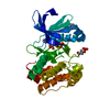 5os0C  5os1C 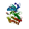 5os3C  5os4C 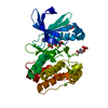 5os5C  5os6C 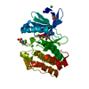 5osdC  5oseC  5osfC C: 同じ文献を引用 ( |
|---|---|
| 類似構造データ |
- リンク
リンク
- 集合体
集合体
| 登録構造単位 | 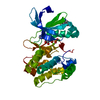
| ||||||||
|---|---|---|---|---|---|---|---|---|---|
| 1 |
| ||||||||
| 単位格子 |
|
- 要素
要素
| #1: タンパク質 | 分子量: 30718.256 Da / 分子数: 1 / 由来タイプ: 組換発現 / 由来: (組換発現)  Homo sapiens (ヒト) Homo sapiens (ヒト)遺伝子: AURKA, AIK, AIRK1, ARK1, AURA, AYK1, BTAK, IAK1, STK15, STK6 発現宿主:  参照: UniProt: O14965, non-specific serine/threonine protein kinase | ||||
|---|---|---|---|---|---|
| #2: 化合物 | ChemComp-ADP / | ||||
| #3: 化合物 | | #4: 化合物 | ChemComp-A7K / [ | #5: 水 | ChemComp-HOH / | |
-実験情報
-実験
| 実験 | 手法:  X線回折 / 使用した結晶の数: 1 X線回折 / 使用した結晶の数: 1 |
|---|
- 試料調製
試料調製
| 結晶 | マシュー密度: 2.78 Å3/Da / 溶媒含有率: 55.78 % |
|---|---|
| 結晶化 | 温度: 298 K / 手法: 蒸気拡散法, シッティングドロップ法 / pH: 8.5 詳細: 0.1 M Tris, pH 8.5: 0.5 M NaCl: 0.2 M MgCl2: 32.5 % v/v PEG 3350 |
-データ収集
| 回折 | 平均測定温度: 100 K | ||||||||||||||||||||||||||||||||||||||||||||||||||||||||||||||||||||||||||||||||||||||||||||||||||||||||||||||||||||||||||||||||||||||||||||||||||||||||||||||||||||||||
|---|---|---|---|---|---|---|---|---|---|---|---|---|---|---|---|---|---|---|---|---|---|---|---|---|---|---|---|---|---|---|---|---|---|---|---|---|---|---|---|---|---|---|---|---|---|---|---|---|---|---|---|---|---|---|---|---|---|---|---|---|---|---|---|---|---|---|---|---|---|---|---|---|---|---|---|---|---|---|---|---|---|---|---|---|---|---|---|---|---|---|---|---|---|---|---|---|---|---|---|---|---|---|---|---|---|---|---|---|---|---|---|---|---|---|---|---|---|---|---|---|---|---|---|---|---|---|---|---|---|---|---|---|---|---|---|---|---|---|---|---|---|---|---|---|---|---|---|---|---|---|---|---|---|---|---|---|---|---|---|---|---|---|---|---|---|---|---|---|---|
| 放射光源 | 由来:  シンクロトロン / サイト: シンクロトロン / サイト:  Diamond Diamond  / ビームライン: I04-1 / 波長: 0.9282 Å / ビームライン: I04-1 / 波長: 0.9282 Å | ||||||||||||||||||||||||||||||||||||||||||||||||||||||||||||||||||||||||||||||||||||||||||||||||||||||||||||||||||||||||||||||||||||||||||||||||||||||||||||||||||||||||
| 検出器 | タイプ: DECTRIS PILATUS 6M-F / 検出器: PIXEL / 日付: 2016年2月15日 | ||||||||||||||||||||||||||||||||||||||||||||||||||||||||||||||||||||||||||||||||||||||||||||||||||||||||||||||||||||||||||||||||||||||||||||||||||||||||||||||||||||||||
| 放射 | プロトコル: SINGLE WAVELENGTH / 単色(M)・ラウエ(L): M / 散乱光タイプ: x-ray | ||||||||||||||||||||||||||||||||||||||||||||||||||||||||||||||||||||||||||||||||||||||||||||||||||||||||||||||||||||||||||||||||||||||||||||||||||||||||||||||||||||||||
| 放射波長 | 波長: 0.9282 Å / 相対比: 1 | ||||||||||||||||||||||||||||||||||||||||||||||||||||||||||||||||||||||||||||||||||||||||||||||||||||||||||||||||||||||||||||||||||||||||||||||||||||||||||||||||||||||||
| 反射 | 解像度: 1.92→65.622 Å / Num. obs: 49856 / % possible obs: 99.9 % / Observed criterion σ(I): -3 / 冗長度: 10.306 % / Biso Wilson estimate: 46.986 Å2 / CC1/2: 0.999 / Rmerge(I) obs: 0.077 / Rrim(I) all: 0.081 / Χ2: 0.96 / Net I/σ(I): 16.17 | ||||||||||||||||||||||||||||||||||||||||||||||||||||||||||||||||||||||||||||||||||||||||||||||||||||||||||||||||||||||||||||||||||||||||||||||||||||||||||||||||||||||||
| 反射 シェル | Diffraction-ID: 1
|
- 解析
解析
| ソフトウェア |
| ||||||||||||||||||||||||
|---|---|---|---|---|---|---|---|---|---|---|---|---|---|---|---|---|---|---|---|---|---|---|---|---|---|
| 精密化 | 解像度: 1.92→65.622 Å / SU ML: 0.3 / 交差検証法: FREE R-VALUE / σ(F): 1.35 / 位相誤差: 25.51
| ||||||||||||||||||||||||
| 溶媒の処理 | 減衰半径: 0.9 Å / VDWプローブ半径: 1.11 Å | ||||||||||||||||||||||||
| 原子変位パラメータ | Biso max: 113.5 Å2 / Biso mean: 39.3632 Å2 / Biso min: 21.42 Å2 | ||||||||||||||||||||||||
| 精密化ステップ | サイクル: final / 解像度: 1.92→65.622 Å
|
 ムービー
ムービー コントローラー










 PDBj
PDBj


