| Entry | Database: PDB / ID: 5or1
|
|---|
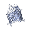
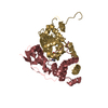
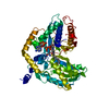
| Title | BamA structure of Salmonella enterica |
|---|
 Components Components | Outer membrane protein assembly factor BamA |
|---|
 Keywords Keywords | MEMBRANE PROTEIN / Outer membrane protein |
|---|
| Function / homology |  Function and homology information Function and homology information
Bam protein complex / Gram-negative-bacterium-type cell outer membrane assembly / protein insertion into membraneSimilarity search - Function membrane protein fhac: a member of the omp85/tpsb transporter family / Outer membrane protein assembly factor BamA / POTRA domain, BamA/TamA-like / Surface antigen variable number repeat / Surface antigen D15-like / POTRA domain / POTRA domain profile. / Bacterial surface antigen (D15) / Omp85 superfamily domain / Porin ...membrane protein fhac: a member of the omp85/tpsb transporter family / Outer membrane protein assembly factor BamA / POTRA domain, BamA/TamA-like / Surface antigen variable number repeat / Surface antigen D15-like / POTRA domain / POTRA domain profile. / Bacterial surface antigen (D15) / Omp85 superfamily domain / Porin / Beta Barrel / Mainly BetaSimilarity search - Domain/homology |
|---|
| Biological species |  Salmonella typhimurium (bacteria) Salmonella typhimurium (bacteria) |
|---|
| Method |  X-RAY DIFFRACTION / SYNCHROTRON / MOLECULAR REPLACEMENT / Resolution: 2.92 Å X-RAY DIFFRACTION / SYNCHROTRON / MOLECULAR REPLACEMENT / Resolution: 2.92 Å |
|---|
 Authors Authors | Dong, C. / Gu, Y. |
|---|
 Citation Citation | Journal: Biochem. J. / Year: 2017
Title: BamA beta 16C strand and periplasmic turns are critical for outer membrane protein insertion and assembly.
Authors: Gu, Y. / Zeng, Y. / Wang, Z. / Dong, C. |
|---|
| History | | Deposition | Aug 14, 2017 | Deposition site: PDBE / Processing site: PDBE |
|---|
| Revision 1.0 | Feb 14, 2018 | Provider: repository / Type: Initial release |
|---|
| Revision 1.1 | Oct 16, 2019 | Group: Data collection / Category: reflns_shell |
|---|
| Revision 1.2 | May 8, 2024 | Group: Data collection / Database references / Category: chem_comp_atom / chem_comp_bond / database_2
Item: _database_2.pdbx_DOI / _database_2.pdbx_database_accession |
|---|
|
|---|
 Movie
Movie Controller


 Open data
 Basic information
 Structure visualization
 Downloads & links
 Other downloads
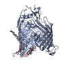


 PDBj
PDBj Assembly
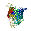
 Sample preparation
 / Beamline: I04-1 / Wavelength: 0.92 Å
 Processing