- PDB-5om9: Crystal structure of the human CARBOXYPEPTIDASE A1 in complex wit... -
+
Open data
ID or keywords:
Loading...
-
Basic information
Entry
Database: PDB / ID: 5om9
Title
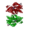
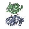
Crystal structure of the human CARBOXYPEPTIDASE A1 in complex with a thiirane mechanism-based inhibitor
Components
Carboxypeptidase A1
Keywords
HYDROLASE
Function / homology
Function and homology information
carboxypeptidase A / Developmental Lineage of Pancreatic Acinar Cells / leukotriene metabolic process / response to cadmium ion / metallocarboxypeptidase activity / : / proteolysis / : / zinc ion binding Similarity search - Function
Mass: 47190.027 Da / Num. of mol.: 2 Source method: isolated from a genetically manipulated source Source: (gene. exp.) Homo sapiens (human) / Gene: CPA1, CPA / Production host: Escherichia coli (E. coli) / References: UniProt: P15085, carboxypeptidase A
Resolution: 1.8→43.68 Å / Cor.coef. Fo:Fc: 0.958 / Cor.coef. Fo:Fc free: 0.921 / SU B: 5.357 / SU ML: 0.077 / Cross valid method: THROUGHOUT / ESU R: 0.465 / ESU R Free: 0.128 / Details: HYDROGENS HAVE BEEN ADDED IN THE RIDING POSITIONS
Rfactor
Num. reflection
% reflection
Selection details
Rfree
0.21178
3390
5 %
RANDOM
Rwork
0.15445
-
-
-
obs
0.15732
63800
96.48 %
-
Solvent computation
Ion probe radii: 0.8 Å / Shrinkage radii: 0.8 Å / VDW probe radii: 1.4 Å
 Movie
Movie Controller

 Yorodumi
 Open data
 Basic information
 Components
 Keywords
 Function and homology information
 Homo sapiens (human)
 X-RAY DIFFRACTION /
  Authors
 Citation
 Structure visualization
 Downloads & links
 Other downloads


 PDBj
PDBj
 Assembly
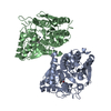

 Mass: 176.276 Da / Num. of mol.: 2 / Source method: obtained synthetically / Formula: C8H16O2S
 Mass: 65.409 Da / Num. of mol.: 3 / Source method: obtained synthetically / Formula: Zn
 Mass: 18.015 Da / Num. of mol.: 443 / Source method: isolated from a natural source / Formula: H2O
 Sample preparation
 / Beamline: XALOC / Wavelength: 0.97949 Å
 Processing