 Movie
Movie Controller

[English] 日本語
 Yorodumi
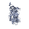
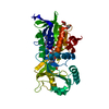
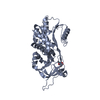
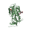
- PDB-5om6: Crystal structure of Alpha1-antichymotrypsin variant DBS-I-allo2:... -
+ Open data

- Basic information
| Entry | Database: PDB / ID: 5om6 | ||||||
|---|---|---|---|---|---|---|---|
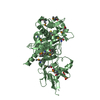
| Title | Crystal structure of Alpha1-antichymotrypsin variant DBS-I-allo2: a MMP9-cleavable drug-binding serpin for doxycycline | ||||||
 Components Components | (Alpha-1-antichymotrypsin) x 2 | ||||||
 Keywords Keywords | TRANSPORT PROTEIN / Serpin / alpha1-Antichymotrypsin / Doxycycline-binding protein / MMP9-cleavable RCL | ||||||
| Function / homology |  Function and homology information Function and homology informationmaintenance of gastrointestinal epithelium / regulation of lipid metabolic process / response to cytokine / platelet alpha granule lumen / acute-phase response / serine-type endopeptidase inhibitor activity / azurophil granule lumen / Platelet degranulation / extracellular matrix / secretory granule lumen ...maintenance of gastrointestinal epithelium / regulation of lipid metabolic process / response to cytokine / platelet alpha granule lumen / acute-phase response / serine-type endopeptidase inhibitor activity / azurophil granule lumen / Platelet degranulation / extracellular matrix / secretory granule lumen / blood microparticle / inflammatory response / Neutrophil degranulation / : / DNA binding / extracellular exosome / extracellular region / nucleus Similarity search - Function | ||||||
| Biological species |  Homo sapiens (human) Homo sapiens (human) | ||||||
| Method |  X-RAY DIFFRACTION / SYNCHROTRON / MOLECULAR REPLACEMENT / Resolution: 1.849 Å X-RAY DIFFRACTION / SYNCHROTRON / MOLECULAR REPLACEMENT / Resolution: 1.849 Å | ||||||
 Authors Authors | Schmidt, K. / Muller, Y.A. | ||||||
| Funding support |  Germany, 1items Germany, 1items
| ||||||
 Citation Citation | Journal: Proc. Natl. Acad. Sci. U.S.A. / Year: 2018 Title: Design of an allosterically modulated doxycycline and doxorubicin drug-binding protein. Authors: Schmidt, K. / Gardill, B.R. / Kern, A. / Kirchweger, P. / Borsch, M. / Muller, Y.A. | ||||||
| History |
|
- Structure visualization
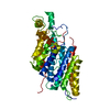
| Structure viewer | Molecule: MolmilJmol/JSmol |
|---|
- Downloads & links
-Download
| PDBx/mmCIF format | 5om6.cif.gz | 172.6 KB | Display | PDBx/mmCIF format |
|---|---|---|---|---|
| PDB format | pdb5om6.ent.gz | 135 KB | Display | PDB format |
| PDBx/mmJSON format | 5om6.json.gz | Tree view | PDBx/mmJSON format | |
| Others |  Other downloads Other downloads |
-Validation report
| Arichive directory | https://data.pdbj.org/pub/pdb/validation_reports/om/5om6ftp://data.pdbj.org/pub/pdb/validation_reports/om/5om6 | HTTPS FTP |
|---|
-Related structure data
| Related structure data | 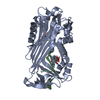 5om2C 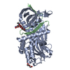 5om3C  5om5C  5om7C 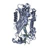 5om8C  6ftpC C: citing same article ( |
|---|---|
| Similar structure data |

-Links
 PDBj
PDBj
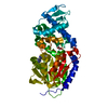
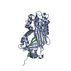
- Assembly
| Deposited unit | 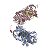
| ||||||||
|---|---|---|---|---|---|---|---|---|---|
| 1 | 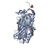
| ||||||||
| 2 | 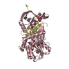
| ||||||||
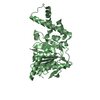
| Unit cell |
|
-Components
-Protein / Protein/peptide , 2 types, 4 molecules ACBD
| #1: Protein | Mass: 41924.457 Da / Num. of mol.: 2 / Fragment: UNP residues 36-383 Mutation: L24R W194F W215Y E242Q K244N L269S P270Q K274S W276F R277F A349R V355L I357G T358P L359R L360Q Source method: isolated from a genetically manipulated source Details: N-terminal residues that are present in the sample sequence but not in the PDB file could not be modelled due to missing electron density. C-terminal residues (ITA...KQA) are in chain I and K Source: (gene. exp.) Homo sapiens (human) / Gene: SERPINA3, AACT, GIG24, GIG25 / Production host:  #2: Protein/peptide | Mass: 4762.639 Da / Num. of mol.: 2 / Fragment: UNP residues 384-423 Mutation: L24R W194F W215Y E242Q K244N L269S P270Q K274S W276F R277F A349R V355L I357G T358P L359R L360Q Source method: isolated from a genetically manipulated source Source: (gene. exp.) Homo sapiens (human) / Gene: SERPINA3, AACT, GIG24, GIG25 / Production host: |
|---|
-Non-polymers , 4 types, 379 molecules 


| #3: Chemical |  Mass: 192.124 Da / Num. of mol.: 2 / Source method: obtained synthetically / Formula: C6H8O7 Mass: 192.124 Da / Num. of mol.: 2 / Source method: obtained synthetically / Formula: C6H8O7#4: Chemical |  Mass: 62.068 Da / Num. of mol.: 2 / Source method: obtained synthetically / Formula: C2H6O2 Mass: 62.068 Da / Num. of mol.: 2 / Source method: obtained synthetically / Formula: C2H6O2#5: Chemical | ChemComp-CL / |  Mass: 35.453 Da / Num. of mol.: 1 / Source method: obtained synthetically / Formula: Cl Mass: 35.453 Da / Num. of mol.: 1 / Source method: obtained synthetically / Formula: Cl#6: Water | ChemComp-HOH / | Mass: 18.015 Da / Num. of mol.: 374 / Source method: isolated from a natural source / Formula: H2O |
|---|
-Experimental details
-Experiment
| Experiment | Method: X-RAY DIFFRACTION / Number of used crystals: 1 |
|---|
- Sample preparation
| Crystal | Density Matthews: 2.32 Å3/Da / Density % sol: 46.9 % |
|---|---|
| Crystal grow | Temperature: 293 K / Method: vapor diffusion, sitting drop / Details: 0.1 M citric acid pH 3.5, 25 % w/v PEG 3350 |
-Data collection
| Diffraction | Mean temperature: 100 K |
|---|---|
| Diffraction source | Source: SYNCHROTRON / Site: BESSY / Beamline: 14.1 / Wavelength: 0.9184 Å |
| Detector | Type: DECTRIS PILATUS3 S 6M / Detector: PIXEL / Date: Sep 15, 2016 |
| Radiation | Protocol: SINGLE WAVELENGTH / Monochromatic (M) / Laue (L): M / Scattering type: x-ray |
| Radiation wavelength | Wavelength: 0.9184 Å / Relative weight: 1 |
| Reflection | Resolution: 1.849→38.601 Å / Num. obs: 72070 / % possible obs: 98.7 % / Redundancy: 3.8 % / Biso Wilson estimate: 33.2 Å2 / CC1/2: 0.999 / Rrim(I) all: 0.068 / Net I/σ(I): 15.2 |
| Reflection shell | Resolution: 1.85→1.96 Å / Redundancy: 3.7 % / Mean I/σ(I) obs: 2.1 / Num. unique obs: 11566 / CC1/2: 0.766 / Rrim(I) all: 0.748 / % possible all: 98.4 |
- Processing
| Software |
| ||||||||||||||||||||||||||||||||||||||||||||||||||||||||||||||||||||||||||||||||||||||||||||||||||||||||||||||||
|---|---|---|---|---|---|---|---|---|---|---|---|---|---|---|---|---|---|---|---|---|---|---|---|---|---|---|---|---|---|---|---|---|---|---|---|---|---|---|---|---|---|---|---|---|---|---|---|---|---|---|---|---|---|---|---|---|---|---|---|---|---|---|---|---|---|---|---|---|---|---|---|---|---|---|---|---|---|---|---|---|---|---|---|---|---|---|---|---|---|---|---|---|---|---|---|---|---|---|---|---|---|---|---|---|---|---|---|---|---|---|---|---|---|
| Refinement | Method to determine structure: MOLECULAR REPLACEMENT / Resolution: 1.849→38.601 Å / SU ML: 0.25 / Cross valid method: FREE R-VALUE / σ(F): 1.35 / Phase error: 27.61
| ||||||||||||||||||||||||||||||||||||||||||||||||||||||||||||||||||||||||||||||||||||||||||||||||||||||||||||||||
| Solvent computation | Shrinkage radii: 0.9 Å / VDW probe radii: 1.11 Å | ||||||||||||||||||||||||||||||||||||||||||||||||||||||||||||||||||||||||||||||||||||||||||||||||||||||||||||||||
| Refinement step | Cycle: LAST / Resolution: 1.849→38.601 Å
| ||||||||||||||||||||||||||||||||||||||||||||||||||||||||||||||||||||||||||||||||||||||||||||||||||||||||||||||||
| Refine LS restraints |
| ||||||||||||||||||||||||||||||||||||||||||||||||||||||||||||||||||||||||||||||||||||||||||||||||||||||||||||||||
| LS refinement shell |
|
