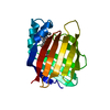
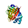
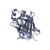
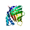
Entry Database : PDB / ID : 5ogbTitle Human Cellular Retinoic Acid Binding Protein II (CRABPII) with bound synthetic retinoid DC360. Cellular retinoic acid-binding protein 2 Keywords / / / / Function / homology Function Domain/homology Component
/ / / / / / / / / / / / / / / / / / / / / / / / / / / / / / / / / / / / / Biological species Homo sapiens (human)Method / / / Resolution : 1.8 Å Authors Chisholm, D. / Tomlinson, C. / Whiting, A. / Pohl, E. Funding support Organization Grant number Country Medical Research Council (United Kingdom)
Journal : Acs Chem.Biol. / Year : 2019Title : Fluorescent Retinoic Acid Analogues as Probes for Biochemical and Intracellular Characterization of Retinoid Signaling Pathways.Authors : Chisholm, D.R. / Tomlinson, C.W.E. / Zhou, G.L. / Holden, C. / Affleck, V. / Lamb, R. / Newling, K. / Ashton, P. / Valentine, R. / Redfern, C. / Erostyak, J. / Makkai, G. / Ambler, C.A. / Whiting, A. / Pohl, E. History Deposition Jul 12, 2017 Deposition site / Processing site Revision 1.0 Oct 24, 2018 Provider / Type Revision 1.1 May 8, 2019 Group / Database references / Category / citation_author / pdbx_database_procItem _citation.country / _citation.journal_abbrev ... _citation.country / _citation.journal_abbrev / _citation.journal_id_CSD / _citation.journal_id_ISSN / _citation.journal_volume / _citation.page_first / _citation.page_last / _citation.pdbx_database_id_DOI / _citation.pdbx_database_id_PubMed / _citation.title / _citation.year Revision 1.2 Oct 16, 2019 Group / Category Revision 1.3 Jan 17, 2024 Group / Database references / Refinement descriptionCategory chem_comp_atom / chem_comp_bond ... chem_comp_atom / chem_comp_bond / database_2 / pdbx_initial_refinement_model Item / _database_2.pdbx_database_accession
Show all Show less
 Movie
Movie Controller

 Yorodumi
 Open data
 Basic information
 Components
 Keywords
 Function and homology information
 Homo sapiens (human)
 X-RAY DIFFRACTION /
  Authors
 United Kingdom, 1items
  Citation
 Structure visualization
 Downloads & links
 Other downloads
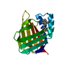


 PDBj
PDBj


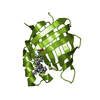
 Assembly
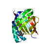


 Mass: 345.434 Da / Num. of mol.: 1 / Source method: obtained synthetically / Formula: C23H23NO2
 Mass: 18.015 Da / Num. of mol.: 62 / Source method: isolated from a natural source / Formula: H2O
 Sample preparation
 Processing