 Movie
Movie Controller

[English] 日本語
 Yorodumi
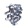
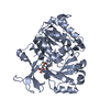
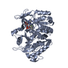
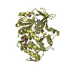
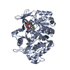
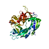
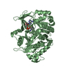
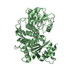
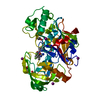
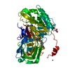
- PDB-5oe5: Crystal structure of the N-terminal domain of PqsA in complex wit... -
+ Open data

- Basic information
| Entry | Database: PDB / ID: 5oe5 | ||||||
|---|---|---|---|---|---|---|---|
| Title | Crystal structure of the N-terminal domain of PqsA in complex with anthraniloyl-AMP (crystal form 3) | ||||||
 Components Components | Anthranilate--CoA ligase | ||||||
 Keywords Keywords | LIGASE / PQS / PqsA / Anthranilate / Anthraniloyl-AMP / Anthraniloyl-CoA / Pseudomonas Quinolone Signal / Pseudomonas aeruginosa / Quorum Sensing / Aryl-CoA ligase / ANL superfamily | ||||||
| Function / homology |  Function and homology information Function and homology informationanthranilate-CoA ligase / anthranilate-CoA ligase activity / acid-thiol ligase activity / secondary metabolite biosynthetic process / ATP binding Similarity search - Function | ||||||
| Biological species |  Pseudomonas aeruginosa PAO1 (bacteria) Pseudomonas aeruginosa PAO1 (bacteria) | ||||||
| Method |  X-RAY DIFFRACTION / SYNCHROTRON / MOLECULAR REPLACEMENT / molecular replacement / Resolution: 1.74 Å X-RAY DIFFRACTION / SYNCHROTRON / MOLECULAR REPLACEMENT / molecular replacement / Resolution: 1.74 Å | ||||||
 Authors Authors | Witzgall, F. / Ewert, W. / Blankenfeldt, W. | ||||||
 Citation Citation | Journal: Chembiochem / Year: 2017 Title: Structures of the N-Terminal Domain of PqsA in Complex with Anthraniloyl- and 6-Fluoroanthraniloyl-AMP: Substrate Activation in Pseudomonas Quinolone Signal (PQS) Biosynthesis. Authors: Witzgall, F. / Ewert, W. / Blankenfeldt, W. | ||||||
| History |
|
- Structure visualization
| Structure viewer | Molecule: MolmilJmol/JSmol |
|---|
- Downloads & links
-Download
| PDBx/mmCIF format | 5oe5.cif.gz | 240.9 KB | Display | PDBx/mmCIF format |
|---|---|---|---|---|
| PDB format | pdb5oe5.ent.gz | 197.1 KB | Display | PDB format |
| PDBx/mmJSON format | 5oe5.json.gz | Tree view | PDBx/mmJSON format | |
| Others |  Other downloads Other downloads |
-Validation report
| Arichive directory | https://data.pdbj.org/pub/pdb/validation_reports/oe/5oe5ftp://data.pdbj.org/pub/pdb/validation_reports/oe/5oe5 | HTTPS FTP |
|---|
-Related structure data


-Links
 PDBj
PDBj
- Assembly
| Deposited unit | 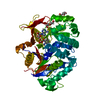
| ||||||||
|---|---|---|---|---|---|---|---|---|---|
| 1 |
| ||||||||
| Unit cell |
|
-Components
| #1: Protein | Mass: 44395.195 Da / Num. of mol.: 1 Source method: isolated from a genetically manipulated source Source: (gene. exp.) Pseudomonas aeruginosa PAO1 (bacteria) / Gene: pqsA, PA0996 / Production host: | ||
|---|---|---|---|
| #2: Chemical | ChemComp-3UK / 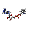  Mass: 466.342 Da / Num. of mol.: 1 / Source method: obtained synthetically / Formula: C17H19N6O8P Mass: 466.342 Da / Num. of mol.: 1 / Source method: obtained synthetically / Formula: C17H19N6O8P | ||
| #3: Chemical |   Mass: 106.120 Da / Num. of mol.: 2 / Source method: obtained synthetically / Formula: C4H10O3 Mass: 106.120 Da / Num. of mol.: 2 / Source method: obtained synthetically / Formula: C4H10O3#4: Water | ChemComp-HOH / |  Mass: 18.015 Da / Num. of mol.: 329 / Source method: isolated from a natural source / Formula: H2O Mass: 18.015 Da / Num. of mol.: 329 / Source method: isolated from a natural source / Formula: H2O |
-Experimental details
-Experiment
| Experiment | Method: X-RAY DIFFRACTION / Number of used crystals: 1 |
|---|
- Sample preparation
| Crystal | Density Matthews: 2.12 Å3/Da / Density % sol: 42.06 % |
|---|---|
| Crystal grow | Temperature: 293 K / Method: vapor diffusion, sitting drop / pH: 6.3 Details: 100 mM tri-sodium citrate (pH 6.3), 240 mM ammonium acetate, 28.5% PEG 3350 |
-Data collection
| Diffraction | Mean temperature: 100 K | |||||||||||||||||||||
|---|---|---|---|---|---|---|---|---|---|---|---|---|---|---|---|---|---|---|---|---|---|---|
| Diffraction source | Source: SYNCHROTRON / Site: SLS  / Beamline: X06DA / Wavelength: 1 Å / Beamline: X06DA / Wavelength: 1 Å | |||||||||||||||||||||
| Detector | Type: DECTRIS PILATUS 2M-F / Detector: PIXEL / Date: Feb 23, 2016 | |||||||||||||||||||||
| Radiation | Protocol: SINGLE WAVELENGTH / Monochromatic (M) / Laue (L): M / Scattering type: x-ray | |||||||||||||||||||||
| Radiation wavelength | Wavelength: 1 Å / Relative weight: 1 | |||||||||||||||||||||
| Reflection | Resolution: 1.74→47.24 Å / Num. obs: 39169 / % possible obs: 99.3 % / Redundancy: 12.6 % / Biso Wilson estimate: 18.34 Å2 / CC1/2: 0.995 / Rmerge(I) obs: 0.185 / Rpim(I) all: 0.054 / Rrim(I) all: 0.193 / Net I/σ(I): 10.2 | |||||||||||||||||||||
| Reflection shell | Diffraction-ID: 1
|
-Phasing
| Phasing | Method: molecular replacement |
|---|
- Processing
| Software |
| ||||||||||||||||||||||||||||||||||||||||||||||||||||||||||||||||||||||||||||||||||||||||||||||||||||
|---|---|---|---|---|---|---|---|---|---|---|---|---|---|---|---|---|---|---|---|---|---|---|---|---|---|---|---|---|---|---|---|---|---|---|---|---|---|---|---|---|---|---|---|---|---|---|---|---|---|---|---|---|---|---|---|---|---|---|---|---|---|---|---|---|---|---|---|---|---|---|---|---|---|---|---|---|---|---|---|---|---|---|---|---|---|---|---|---|---|---|---|---|---|---|---|---|---|---|---|---|---|
| Refinement | Method to determine structure: MOLECULAR REPLACEMENT / Resolution: 1.74→47.236 Å / SU ML: 0.27 / Cross valid method: FREE R-VALUE / σ(F): 1.35 / Phase error: 27.43
| ||||||||||||||||||||||||||||||||||||||||||||||||||||||||||||||||||||||||||||||||||||||||||||||||||||
| Solvent computation | Shrinkage radii: 0.9 Å / VDW probe radii: 1.11 Å | ||||||||||||||||||||||||||||||||||||||||||||||||||||||||||||||||||||||||||||||||||||||||||||||||||||
| Displacement parameters | Biso max: 88.88 Å2 / Biso mean: 27.252 Å2 / Biso min: 7.23 Å2 | ||||||||||||||||||||||||||||||||||||||||||||||||||||||||||||||||||||||||||||||||||||||||||||||||||||
| Refinement step | Cycle: final / Resolution: 1.74→47.236 Å
| ||||||||||||||||||||||||||||||||||||||||||||||||||||||||||||||||||||||||||||||||||||||||||||||||||||
| Refine LS restraints |
| ||||||||||||||||||||||||||||||||||||||||||||||||||||||||||||||||||||||||||||||||||||||||||||||||||||
| LS refinement shell | Refine-ID: X-RAY DIFFRACTION / Rfactor Rfree error: 0 / Total num. of bins used: 13
| ||||||||||||||||||||||||||||||||||||||||||||||||||||||||||||||||||||||||||||||||||||||||||||||||||||
| Refinement TLS params. | Method: refined / Refine-ID: X-RAY DIFFRACTION
| ||||||||||||||||||||||||||||||||||||||||||||||||||||||||||||||||||||||||||||||||||||||||||||||||||||
| Refinement TLS group |
|
