 Movie
Movie Controller

[English] 日本語
 Yorodumi
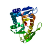
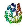
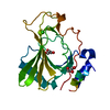
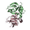
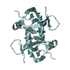
- PDB-5oc6: Crystal structure of human tRNA-dihydrouridine(20) synthase dsRBD... -
+ Open data

- Basic information
| Entry | Database: PDB / ID: 5oc6 | ||||||
|---|---|---|---|---|---|---|---|
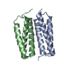
| Title | Crystal structure of human tRNA-dihydrouridine(20) synthase dsRBD in complex with a 22 nucleotide dsRNA | ||||||
 Components Components |
| ||||||
 Keywords Keywords | RNA BINDING PROTEIN / double-stranded RNA-binding domain protein-RNA complex dsRNA | ||||||
| Function / homology |  Function and homology information Function and homology informationtRNA-dihydrouridine20 synthase [NAD(P)+] / tRNA-dihydrouridine20 synthase activity / tRNA dihydrouridine synthesis / tRNA dihydrouridine synthase activity / tRNA modification in the nucleus and cytosol / protein kinase inhibitor activity / NADPH binding / antiviral innate immune response / PKR-mediated signaling / FMN binding ...tRNA-dihydrouridine20 synthase [NAD(P)+] / tRNA-dihydrouridine20 synthase activity / tRNA dihydrouridine synthesis / tRNA dihydrouridine synthase activity / tRNA modification in the nucleus and cytosol / protein kinase inhibitor activity / NADPH binding / antiviral innate immune response / PKR-mediated signaling / FMN binding / flavin adenine dinucleotide binding / double-stranded RNA binding / tRNA binding / endoplasmic reticulum / mitochondrion / cytoplasm / cytosol Similarity search - Function | ||||||
| Biological species |  Homo sapiens (human) Homo sapiens (human)synthetic construct (others) | ||||||
| Method |  X-RAY DIFFRACTION / SYNCHROTRON / MOLECULAR REPLACEMENT / Resolution: 3.2 Å X-RAY DIFFRACTION / SYNCHROTRON / MOLECULAR REPLACEMENT / Resolution: 3.2 Å | ||||||
 Authors Authors | Bou-nader, C. / Pecqueur, L. / Hamdane, D. | ||||||
 Citation Citation | Journal: Nucleic Acids Res. / Year: 2019 Title: Molecular basis for transfer RNA recognition by the double-stranded RNA-binding domain of human dihydrouridine synthase 2. Authors: Bou-Nader, C. / Barraud, P. / Pecqueur, L. / Perez, J. / Velours, C. / Shepard, W. / Fontecave, M. / Tisne, C. / Hamdane, D. | ||||||
| History |
|
- Structure visualization
| Structure viewer | Molecule: MolmilJmol/JSmol |
|---|
- Downloads & links
-Download
| PDBx/mmCIF format | 5oc6.cif.gz | 77.1 KB | Display | PDBx/mmCIF format |
|---|---|---|---|---|
| PDB format | pdb5oc6.ent.gz | 55.9 KB | Display | PDB format |
| PDBx/mmJSON format | 5oc6.json.gz | Tree view | PDBx/mmJSON format | |
| Others |  Other downloads Other downloads |
-Validation report
| Arichive directory | https://data.pdbj.org/pub/pdb/validation_reports/oc/5oc6ftp://data.pdbj.org/pub/pdb/validation_reports/oc/5oc6 | HTTPS FTP |
|---|
-Related structure data
| Related structure data |  5oc4C 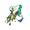 5oc5C 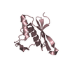 4wftS S: Starting model for refinement C: citing same article ( |
|---|---|
| Similar structure data |


-Links
 PDBj
PDBj


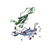
- Assembly
| Deposited unit | 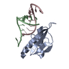
| ||||||||
|---|---|---|---|---|---|---|---|---|---|
| 1 |
| ||||||||
| Unit cell |
|
-Components
| #1: Protein | Mass: 13728.697 Da / Num. of mol.: 1 / Fragment: UNP residues 338-450 Source method: isolated from a genetically manipulated source Source: (gene. exp.) Homo sapiens (human) / Gene: DUS2, DUS2L / Production host:  References: UniProt: Q9NX74, Oxidoreductases; Acting on the CH-CH group of donors; With NAD+ or NADP+ as acceptor |
|---|---|
| #2: RNA chain | Mass: 3482.130 Da / Num. of mol.: 2 / Source method: obtained synthetically / Source: (synth.) synthetic construct (others) |
-Experimental details
-Experiment
| Experiment | Method: X-RAY DIFFRACTION / Number of used crystals: 1 |
|---|
- Sample preparation
| Crystal | Density Matthews: 2.72 Å3/Da / Density % sol: 54.74 % |
|---|---|
| Crystal grow | Temperature: 292 K / Method: vapor diffusion, hanging drop / pH: 6 / Details: 15% v/v isopropanol 20 mM MgCl2 50 mM MES |
-Data collection
| Diffraction | Mean temperature: 100 K |
|---|---|
| Diffraction source | Source: SYNCHROTRON / Site: SOLEIL  / Beamline: PROXIMA 2 / Wavelength: 0.9801 Å / Beamline: PROXIMA 2 / Wavelength: 0.9801 Å |
| Detector | Type: ADSC QUANTUM 315r / Detector: CCD / Date: Jun 13, 2015 |
| Radiation | Protocol: SINGLE WAVELENGTH / Monochromatic (M) / Laue (L): M / Scattering type: x-ray |
| Radiation wavelength | Wavelength: 0.9801 Å / Relative weight: 1 |
| Reflection | Resolution: 3.2→43.65 Å / Num. obs: 3855 / % possible obs: 97.52 % / Redundancy: 5.8 % / Biso Wilson estimate: 100.05 Å2 / CC1/2: 0.995 / Rmerge(I) obs: 0.3064 / Net I/σ(I): 5.11 |
| Reflection shell | Resolution: 3.2→3.314 Å / Redundancy: 3.8 % / Mean I/σ(I) obs: 0.85 / Num. unique obs: 358 / CC1/2: 0.302 / % possible all: 93.44 |
- Processing
| Software |
| ||||||||||||||||||||||||||||||||||||||||||||||||||||||||||||||||||||||||||||||||||||||||||||||||||||||||||||||||||
|---|---|---|---|---|---|---|---|---|---|---|---|---|---|---|---|---|---|---|---|---|---|---|---|---|---|---|---|---|---|---|---|---|---|---|---|---|---|---|---|---|---|---|---|---|---|---|---|---|---|---|---|---|---|---|---|---|---|---|---|---|---|---|---|---|---|---|---|---|---|---|---|---|---|---|---|---|---|---|---|---|---|---|---|---|---|---|---|---|---|---|---|---|---|---|---|---|---|---|---|---|---|---|---|---|---|---|---|---|---|---|---|---|---|---|---|
| Refinement | Method to determine structure: MOLECULAR REPLACEMENT Starting model: 4wft Resolution: 3.2→43.65 Å / Cor.coef. Fo:Fc: 0.926 / Cor.coef. Fo:Fc free: 0.917 / Cross valid method: THROUGHOUT / σ(F): 0 / SU Rfree Blow DPI: 0.438
| ||||||||||||||||||||||||||||||||||||||||||||||||||||||||||||||||||||||||||||||||||||||||||||||||||||||||||||||||||
| Displacement parameters | Biso mean: 99.32 Å2
| ||||||||||||||||||||||||||||||||||||||||||||||||||||||||||||||||||||||||||||||||||||||||||||||||||||||||||||||||||
| Refine analyze | Luzzati coordinate error obs: 0.53 Å | ||||||||||||||||||||||||||||||||||||||||||||||||||||||||||||||||||||||||||||||||||||||||||||||||||||||||||||||||||
| Refinement step | Cycle: 1 / Resolution: 3.2→43.65 Å
| ||||||||||||||||||||||||||||||||||||||||||||||||||||||||||||||||||||||||||||||||||||||||||||||||||||||||||||||||||
| Refine LS restraints |
| ||||||||||||||||||||||||||||||||||||||||||||||||||||||||||||||||||||||||||||||||||||||||||||||||||||||||||||||||||
| LS refinement shell | Resolution: 3.2→3.58 Å / Total num. of bins used: 5
| ||||||||||||||||||||||||||||||||||||||||||||||||||||||||||||||||||||||||||||||||||||||||||||||||||||||||||||||||||
| Refinement TLS params. | Method: refined / Refine-ID: X-RAY DIFFRACTION
| ||||||||||||||||||||||||||||||||||||||||||||||||||||||||||||||||||||||||||||||||||||||||||||||||||||||||||||||||||
| Refinement TLS group |
|
