| 登録情報 | データベース: PDB / ID: 5oak
|
|---|
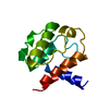
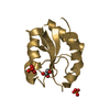
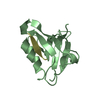
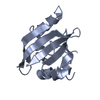
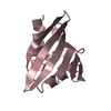
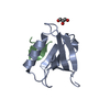
| タイトル | Structure of the dmPar3 PDZ1 domain in complex with the dmPar6 PBM |
|---|
 要素 要素 | Bazooka, isoform C,LD29223p |
|---|
 キーワード キーワード | PROTEIN BINDING / Cell polarity protein |
|---|
| 機能・相同性 |  機能・相同性情報 機能・相同性情報
spot adherens junction / follicle cell of egg chamber-cell adhesion / border follicle cell delamination / germ-band extension / Malpighian tubule development / RAC1 GTPase cycle / RHOV GTPase cycle / terminal branching, open tracheal system / establishment or maintenance of neuroblast polarity / branching involved in open tracheal system development ...spot adherens junction / follicle cell of egg chamber-cell adhesion / border follicle cell delamination / germ-band extension / Malpighian tubule development / RAC1 GTPase cycle / RHOV GTPase cycle / terminal branching, open tracheal system / establishment or maintenance of neuroblast polarity / branching involved in open tracheal system development / zonula adherens assembly / subapical complex / TGF-beta receptor signaling in EMT (epithelial to mesenchymal transition) / photoreceptor cell morphogenesis / establishment of neuroblast polarity / Asymmetric localization of PCP proteins / RHOU GTPase cycle / oocyte anterior/posterior axis specification / oocyte microtubule cytoskeleton organization / muscle cell postsynaptic specialization / germarium-derived oocyte fate determination / asymmetric protein localization involved in cell fate determination / establishment or maintenance of polarity of embryonic epithelium / border follicle cell migration / negative regulation of filopodium assembly / PAR polarity complex / establishment of centrosome localization / asymmetric neuroblast division / establishment of apical/basal cell polarity / apical cortex / zonula adherens / morphogenesis of a polarized epithelium / phosphatidic acid binding / establishment or maintenance of epithelial cell apical/basal polarity / positive regulation of smoothened signaling pathway / apical protein localization / centrosome cycle / positive regulation of filopodium assembly / apical junction complex / establishment of cell polarity / protein kinase inhibitor activity / bicellular tight junction / positive regulation of lamellipodium assembly / synapse assembly / phosphatidylinositol binding / adherens junction / microtubule cytoskeleton organization / terminal bouton / apical part of cell / intracellular protein localization / cell cortex / cell adhesion / apical plasma membrane / plasma membrane類似検索 - 分子機能 Partitioning defective protein 6, PB1 domain / : / Par3/HAL, N-terminal / N-terminal of Par3 and HAL proteins / : / PB1 domain / PB1 domain / PB1 domain / : / PB1 domain profile. ...Partitioning defective protein 6, PB1 domain / : / Par3/HAL, N-terminal / N-terminal of Par3 and HAL proteins / : / PB1 domain / PB1 domain / PB1 domain / : / PB1 domain profile. / PDZ domain / PDZ domain profile. / Domain present in PSD-95, Dlg, and ZO-1/2. / PDZ domain / PDZ superfamily類似検索 - ドメイン・相同性 |
|---|
| 生物種 |   Drosophila melanogaster (キイロショウジョウバエ) Drosophila melanogaster (キイロショウジョウバエ) |
|---|
| 手法 |  X線回折 / シンクロトロン / 分子置換 / 解像度: 1.5 Å X線回折 / シンクロトロン / 分子置換 / 解像度: 1.5 Å |
|---|
 データ登録者 データ登録者 | Bruekner, S.R. / Wiesner, S. |
|---|
| 資金援助 |  ドイツ, 2件 ドイツ, 2件 | 組織 | 認可番号 | 国 |
|---|
| Max-Planck Gesellschaft | | ドイツ | | IMPRS "From molecules to Organisms" | | ドイツ |
|
|---|
 引用 引用 | ジャーナル: Sci Signal / 年: 2018
タイトル: Structural basis for the interaction between the cell polarity proteins Par3 and Par6.
著者: Renschler, F.A. / Bruekner, S.R. / Salomon, P.L. / Mukherjee, A. / Kullmann, L. / Schutz-Stoffregen, M.C. / Henzler, C. / Pawson, T. / Krahn, M.P. / Wiesner, S. |
|---|
| 履歴 | | 登録 | 2017年6月22日 | 登録サイト: PDBE / 処理サイト: PDBE |
|---|
| 改定 1.0 | 2018年2月21日 | Provider: repository / タイプ: Initial release |
|---|
| 改定 1.1 | 2018年2月28日 | Group: Database references / カテゴリ: citation / citation_author
Item: _citation.journal_abbrev / _citation.pdbx_database_id_PubMed ..._citation.journal_abbrev / _citation.pdbx_database_id_PubMed / _citation.title / _citation_author.name |
|---|
| 改定 1.2 | 2024年1月17日 | Group: Data collection / Database references / Refinement description
カテゴリ: chem_comp_atom / chem_comp_bond ...chem_comp_atom / chem_comp_bond / database_2 / pdbx_initial_refinement_model
Item: _database_2.pdbx_DOI / _database_2.pdbx_database_accession |
|---|
|
|---|
 ムービー
ムービー コントローラー


 データを開く
 基本情報
基本情報 構造の表示
構造の表示 ダウンロードとリンク
 その他のダウンロード


 PDBj
PDBj

 集合体
集合体


 分子量: 92.094 Da / 分子数: 1 / 由来タイプ: 合成 / 式: C3H8O3
 分子量: 18.015 Da / 分子数: 144 / 由来タイプ: 天然 / 式: H2O
 試料調製
試料調製 / ビームライン: X10SA / 波長: 1 Å
 解析
解析