 Movie
Movie Controller


+ Open data

- Basic information
| Entry | Database: PDB / ID: 5oab | |||||||||
|---|---|---|---|---|---|---|---|---|---|---|
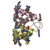
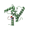
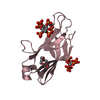
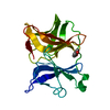
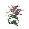
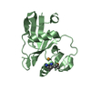
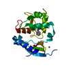
| Title | A novel crystal form of human RNase6 at atomic resolution | |||||||||
 Components Components | Ribonuclease K6 | |||||||||
 Keywords Keywords | HYDROLASE / RNASE K6 / PANCREATIC RIBONUCLEASE | |||||||||
| Function / homology |  Function and homology information Function and homology informationHydrolases; Acting on ester bonds; Endoribonucleases producing 3'-phosphomonoesters / RNA catabolic process / Antimicrobial peptides / RNA nuclease activity / defense response / antimicrobial humoral immune response mediated by antimicrobial peptide / antibacterial humoral response / endonuclease activity / cytoplasmic vesicle / defense response to virus ...Hydrolases; Acting on ester bonds; Endoribonucleases producing 3'-phosphomonoesters / RNA catabolic process / Antimicrobial peptides / RNA nuclease activity / defense response / antimicrobial humoral immune response mediated by antimicrobial peptide / antibacterial humoral response / endonuclease activity / cytoplasmic vesicle / defense response to virus / defense response to Gram-negative bacterium / nucleic acid binding / lysosome / defense response to Gram-positive bacterium / innate immune response / hydrolase activity / : / extracellular region Similarity search - Function | |||||||||
| Biological species |  Homo sapiens (human) Homo sapiens (human) | |||||||||
| Method |  X-RAY DIFFRACTION / SYNCHROTRON / MOLECULAR REPLACEMENT / Resolution: 1.111 Å X-RAY DIFFRACTION / SYNCHROTRON / MOLECULAR REPLACEMENT / Resolution: 1.111 Å | |||||||||
 Authors Authors | Prats-Ejarque, G. / Moussaoui, M. / Boix, E. | |||||||||
| Funding support |  Spain, 2items Spain, 2items
| |||||||||
 Citation Citation | Journal: Biochim Biophys Acta Gen Subj / Year: 2019 Title: Characterization of an RNase with two catalytic centers. Human RNase6 catalytic and phosphate-binding site arrangement favors the endonuclease cleavage of polymeric substrates. Authors: Prats-Ejarque, G. / Blanco, J.A. / Salazar, V.A. / Nogues, V.M. / Moussaoui, M. / Boix, E. #1: Journal: Biochem. J. / Year: 2016Title: The first crystal structure of human RNase 6 reveals a novel substrate-binding and cleavage site arrangement. Authors: Prats-Ejarque, G. / Arranz-Trullen, J. / Blanco, J.A. / Pulido, D. / Nogues, M.V. / Moussaoui, M. / Boix, E. | |||||||||
| History |
|
- Structure visualization
| Structure viewer | Molecule: MolmilJmol/JSmol |
|---|
- Downloads & links
-Download
| PDBx/mmCIF format | 5oab.cif.gz | 119.8 KB | Display | PDBx/mmCIF format |
|---|---|---|---|---|
| PDB format | pdb5oab.ent.gz | 93.2 KB | Display | PDB format |
| PDBx/mmJSON format | 5oab.json.gz | Tree view | PDBx/mmJSON format | |
| Others |  Other downloads Other downloads |
-Validation report
| Arichive directory | https://data.pdbj.org/pub/pdb/validation_reports/oa/5oabftp://data.pdbj.org/pub/pdb/validation_reports/oa/5oab | HTTPS FTP |
|---|
-Related structure data
| Related structure data |  5et4C 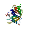 5oghC  6enpC 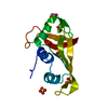 4x09S S: Starting model for refinement C: citing same article ( |
|---|---|
| Similar structure data |


-Links
 PDBj
PDBj


- Assembly
| Deposited unit | 
| |||||||||
|---|---|---|---|---|---|---|---|---|---|---|
| 1 |
| |||||||||
| Unit cell |
| |||||||||
| Components on special symmetry positions |
|
-Components
-Protein , 1 types, 1 molecules A
| #1: Protein | Mass: 14807.069 Da / Num. of mol.: 1 / Fragment: UNP RESIDUES 24-150 Source method: isolated from a genetically manipulated source Source: (gene. exp.) Homo sapiens (human) / Gene: RNASE6, RNS6 / Plasmid: PET11C / Production host:  References: UniProt: Q93091, Hydrolases; Acting on ester bonds; Endoribonucleases producing 3'-phosphomonoesters |
|---|
-Non-polymers , 5 types, 322 molecules 


| #2: Chemical | ChemComp-PO4 /  Mass: 94.971 Da / Num. of mol.: 6 / Source method: obtained synthetically / Formula: PO4 Mass: 94.971 Da / Num. of mol.: 6 / Source method: obtained synthetically / Formula: PO4#3: Chemical |  Mass: 22.990 Da / Num. of mol.: 3 / Source method: obtained synthetically / Formula: Na Mass: 22.990 Da / Num. of mol.: 3 / Source method: obtained synthetically / Formula: Na#4: Chemical |  Mass: 35.453 Da / Num. of mol.: 3 / Source method: obtained synthetically / Formula: Cl Mass: 35.453 Da / Num. of mol.: 3 / Source method: obtained synthetically / Formula: Cl#5: Chemical | ChemComp-K / |  Mass: 39.098 Da / Num. of mol.: 1 / Source method: obtained synthetically / Formula: K Mass: 39.098 Da / Num. of mol.: 1 / Source method: obtained synthetically / Formula: K#6: Water | ChemComp-HOH / | Mass: 18.015 Da / Num. of mol.: 309 / Source method: isolated from a natural source / Formula: H2O |
|---|
-Details
| Has protein modification | Y |
|---|
-Experimental details
-Experiment
| Experiment | Method: X-RAY DIFFRACTION / Number of used crystals: 1 |
|---|
- Sample preparation
| Crystal | Density Matthews: 2.45 Å3/Da / Density % sol: 49.87 % / Description: Cubic |
|---|---|
| Crystal grow | Temperature: 293 K / Method: vapor diffusion, hanging drop / pH: 7 / Details: 1.3 M SODIUM/POTASSIUM PHOSPHATE / PH range: 7-7.5 |
-Data collection
| Diffraction | Mean temperature: 100 K |
|---|---|
| Diffraction source | Source: SYNCHROTRON / Site: ALBA / Beamline: XALOC / Wavelength: 0.9792 Å |
| Detector | Type: DECTRIS PILATUS 6M / Detector: PIXEL / Date: Nov 4, 2016 Details: VERTICAL FOCUSING MIRROR (VFM) AND A HORIZONTAL FOCUSING MIRROR (HFM), MANUFACTURED BY IRELEC |
| Radiation | Monochromator: CHANNEL-CUT DOUBLE CRYSTAL MONOCHROMATOR (CINEL), CRYOCOOLED, 6MM GAP Protocol: SINGLE WAVELENGTH / Monochromatic (M) / Laue (L): M / Scattering type: x-ray |
| Radiation wavelength | Wavelength: 0.9792 Å / Relative weight: 1 |
| Reflection | Resolution: 1.111→47.703 Å / Num. obs: 54593 / % possible obs: 96.2 % / Redundancy: 4 % / Biso Wilson estimate: 9.32 Å2 / CC1/2: 0.996 / Rmerge(I) obs: 0.08221 / Rpim(I) all: 0.04389 / Rrim(I) all: 0.0937 / Net I/σ(I): 9.1 |
| Reflection shell | Resolution: 1.111→1.151 Å / Redundancy: 2.4 % / Rmerge(I) obs: 0.6556 / Mean I/σ(I) obs: 1.47 / Num. unique obs: 4253 / CC1/2: 0.544 / Rpim(I) all: 0.4686 / Rrim(I) all: 0.8125 / % possible all: 75.17 |
- Processing
| Software |
| |||||||||||||||||||||||||||||||||||||||||||||||||||||||||||||||||||||||||||||||||||||||||||||||||||||||||||||||||||||||||||||||||||||
|---|---|---|---|---|---|---|---|---|---|---|---|---|---|---|---|---|---|---|---|---|---|---|---|---|---|---|---|---|---|---|---|---|---|---|---|---|---|---|---|---|---|---|---|---|---|---|---|---|---|---|---|---|---|---|---|---|---|---|---|---|---|---|---|---|---|---|---|---|---|---|---|---|---|---|---|---|---|---|---|---|---|---|---|---|---|---|---|---|---|---|---|---|---|---|---|---|---|---|---|---|---|---|---|---|---|---|---|---|---|---|---|---|---|---|---|---|---|---|---|---|---|---|---|---|---|---|---|---|---|---|---|---|---|---|
| Refinement | Method to determine structure: MOLECULAR REPLACEMENT Starting model: 4X09 Resolution: 1.111→47.703 Å / SU ML: 0.1 / Cross valid method: FREE R-VALUE / σ(F): 1.33 / Phase error: 14.09
| |||||||||||||||||||||||||||||||||||||||||||||||||||||||||||||||||||||||||||||||||||||||||||||||||||||||||||||||||||||||||||||||||||||
| Solvent computation | Shrinkage radii: 0.9 Å / VDW probe radii: 1.11 Å | |||||||||||||||||||||||||||||||||||||||||||||||||||||||||||||||||||||||||||||||||||||||||||||||||||||||||||||||||||||||||||||||||||||
| Displacement parameters | Biso mean: 13.76 Å2 | |||||||||||||||||||||||||||||||||||||||||||||||||||||||||||||||||||||||||||||||||||||||||||||||||||||||||||||||||||||||||||||||||||||
| Refinement step | Cycle: LAST / Resolution: 1.111→47.703 Å
| |||||||||||||||||||||||||||||||||||||||||||||||||||||||||||||||||||||||||||||||||||||||||||||||||||||||||||||||||||||||||||||||||||||
| Refine LS restraints |
| |||||||||||||||||||||||||||||||||||||||||||||||||||||||||||||||||||||||||||||||||||||||||||||||||||||||||||||||||||||||||||||||||||||
| LS refinement shell |
|
