 Movie
Movie Controller

[English] 日本語
 Yorodumi
- PDB-5o8a: Crystal Structure of rsEGFP2 in the non-fluorescent off-state det... -
+ Open data

- Basic information
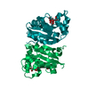
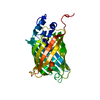
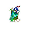
| Entry | Database: PDB / ID: 5o8a | ||||||
|---|---|---|---|---|---|---|---|
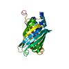
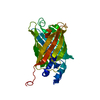
| Title | Crystal Structure of rsEGFP2 in the non-fluorescent off-state determined by SFX | ||||||
 Components Components | Green fluorescent protein | ||||||
 Keywords Keywords | FLUORESCENT PROTEIN / photoswitching on state fluorescent state chromophore SFX serial femtosecond crystallography CXI LCLS time resolved crystallography chromohore isomerisation | ||||||
| Function / homology |  Function and homology information Function and homology informationregulation of viral transcription / symbiont-mediated activation of host NF-kappaB cascade / viral transcription / bioluminescence / transcription antitermination / generation of precursor metabolites and energy / virion component / host cell cytoplasm / host cell nucleus / structural molecule activity ...regulation of viral transcription / symbiont-mediated activation of host NF-kappaB cascade / viral transcription / bioluminescence / transcription antitermination / generation of precursor metabolites and energy / virion component / host cell cytoplasm / host cell nucleus / structural molecule activity / RNA binding / zinc ion binding Similarity search - Function | ||||||
| Biological species |   Aequorea victoria (jellyfish) Aequorea victoria (jellyfish) | ||||||
| Method |  X-RAY DIFFRACTION / FREE ELECTRON LASER / MOLECULAR REPLACEMENT / Resolution: 1.7 Å X-RAY DIFFRACTION / FREE ELECTRON LASER / MOLECULAR REPLACEMENT / Resolution: 1.7 Å | ||||||
 Authors Authors | Coquelle, N. / Sliwa, M. / Woodhouse, J. / Schiro, G. / Adam, V. / Aquila, A. / Barends, T.R.M. / Boutet, S. / Byrdin, M. / Carbajo, S. ...Coquelle, N. / Sliwa, M. / Woodhouse, J. / Schiro, G. / Adam, V. / Aquila, A. / Barends, T.R.M. / Boutet, S. / Byrdin, M. / Carbajo, S. / De la Mora, E. / Doak, R.B. / Feliks, M. / Fieschi, F. / Foucar, L. / Guillon, V. / Hilpert, M. / Hunter, M. / Jakobs, S. / Koglin, J.E. / Kovacsova, G. / Lane, T.J. / Levy, B. / Liang, M. / Nass, K. / Ridard, J. / Robinson, J.S. / Roome, C.M. / Ruckebusch, C. / Seaberg, M. / Thepaut, M. / Cammarata, M. / Demachy, I. / Field, M. / Shoeman, R.L. / Bourgeois, D. / Colletier, J.P. / Schlichting, I. / Weik, M. | ||||||
 Citation Citation | Journal: Nat Chem / Year: 2018 Title: Chromophore twisting in the excited state of a photoswitchable fluorescent protein captured by time-resolved serial femtosecond crystallography. Authors: Coquelle, N. / Sliwa, M. / Woodhouse, J. / Schiro, G. / Adam, V. / Aquila, A. / Barends, T.R.M. / Boutet, S. / Byrdin, M. / Carbajo, S. / De la Mora, E. / Doak, R.B. / Feliks, M. / Fieschi, ...Authors: Coquelle, N. / Sliwa, M. / Woodhouse, J. / Schiro, G. / Adam, V. / Aquila, A. / Barends, T.R.M. / Boutet, S. / Byrdin, M. / Carbajo, S. / De la Mora, E. / Doak, R.B. / Feliks, M. / Fieschi, F. / Foucar, L. / Guillon, V. / Hilpert, M. / Hunter, M.S. / Jakobs, S. / Koglin, J.E. / Kovacsova, G. / Lane, T.J. / Levy, B. / Liang, M. / Nass, K. / Ridard, J. / Robinson, J.S. / Roome, C.M. / Ruckebusch, C. / Seaberg, M. / Thepaut, M. / Cammarata, M. / Demachy, I. / Field, M. / Shoeman, R.L. / Bourgeois, D. / Colletier, J.P. / Schlichting, I. / Weik, M. | ||||||
| History |
|
- Structure visualization
| Structure viewer | Molecule: MolmilJmol/JSmol |
|---|
- Downloads & links
-Download
| PDBx/mmCIF format | 5o8a.cif.gz | 109.7 KB | Display | PDBx/mmCIF format |
|---|---|---|---|---|
| PDB format | pdb5o8a.ent.gz | 87.5 KB | Display | PDB format |
| PDBx/mmJSON format | 5o8a.json.gz | Tree view | PDBx/mmJSON format | |
| Others |  Other downloads Other downloads |
-Validation report
| Arichive directory | https://data.pdbj.org/pub/pdb/validation_reports/o8/5o8aftp://data.pdbj.org/pub/pdb/validation_reports/o8/5o8a | HTTPS FTP |
|---|
-Related structure data
| Related structure data |  5o89C  5o8bC  5o8cC  5dtyS S: Starting model for refinement C: citing same article ( |
|---|---|
| Similar structure data | |
| Experimental dataset #1 | Data reference: 10.11577/1369542 / Data set type: diffraction image data |
| Experimental dataset #2 | Data reference: 10.11577/1369545 / Data set type: diffraction image data |
| Experimental dataset #3 | Data reference: 10.11577/1369546 / Data set type: diffraction image data |
| Experimental dataset #4 | Data reference: 10.11577/1369547 / Data set type: diffraction image data |


-Links
 PDBj
PDBj


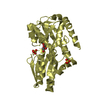
- Assembly
| Deposited unit | 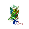
| ||||||||
|---|---|---|---|---|---|---|---|---|---|
| 1 |
| ||||||||
| Unit cell |
|
-Components
| #1: Protein | Mass: 28099.686 Da / Num. of mol.: 1 Source method: isolated from a genetically manipulated source Details: HIS A -8 EXPRESSION TAG HIS A -7 EXPRESSION TAG HIS A -6 EXPRESSION TAG HIS A -5 EXPRESSION TAG HIS A -4 EXPRESSION TAG HIS A -3 EXPRESSION TAG THR A -2 EXPRESSION TAG ASP A -1 EXPRESSION ...Details: HIS A -8 EXPRESSION TAG HIS A -7 EXPRESSION TAG HIS A -6 EXPRESSION TAG HIS A -5 EXPRESSION TAG HIS A -4 EXPRESSION TAG HIS A -3 EXPRESSION TAG THR A -2 EXPRESSION TAG ASP A -1 EXPRESSION TAG PRO A 0 EXPRESSION TAG MET A 1 EXPRESSION TAG VAL A 2 MET 1 ENGINEERED MUTATION LEU A 65 PHE 64 ENGINEERED MUTATION PIA A 68 SER 65 CHROMOPHORE PIA A 68 TYR 66 CHROMOPHORE PIA A 68 GLY 67 CHROMOPHORE LEU A 70 GLN 69 ENGINEERED MUTATION SER A 164 VAL 163 ENGINEERED MUTATION LYS A 207 ALA 206 ENGINEERED MUTATION LEU A 232 HIS 231 ENGINEERED MUTATION Source: (gene. exp.) Aequorea victoria (jellyfish) / Gene: GFP / Production host:  |
|---|---|
| #2: Water | ChemComp-HOH /  Mass: 18.015 Da / Num. of mol.: 311 / Source method: isolated from a natural source / Formula: H2O Mass: 18.015 Da / Num. of mol.: 311 / Source method: isolated from a natural source / Formula: H2O |
-Experimental details
-Experiment
| Experiment | Method: X-RAY DIFFRACTION / Number of used crystals: 1 |
|---|
- Sample preparation
| Crystal | Density Matthews: 2.07 Å3/Da / Density % sol: 40.51 % |
|---|---|
| Crystal grow | Temperature: 293 K / Method: batch mode / pH: 8 / Details: 2 M ammonium sulphate, 20 mM NaCl, 120 mM HEPES |
-Data collection
| Diffraction | Mean temperature: 293 K / Serial crystal experiment: Y |
|---|---|
| Diffraction source | Source: FREE ELECTRON LASER / Site: SLAC LCLS  / Beamline: CXI / Wavelength: 1.3 Å / Beamline: CXI / Wavelength: 1.3 Å |
| Detector | Type: CS-PAD CXI-1 / Detector: PIXEL / Date: May 1, 2015 |
| Radiation | Protocol: SINGLE WAVELENGTH / Monochromatic (M) / Laue (L): M / Scattering type: x-ray |
| Radiation wavelength | Wavelength: 1.3 Å / Relative weight: 1 |
| Reflection | Resolution: 1.7→47.021 Å / Num. obs: 25880 / % possible obs: 99.8 % / Redundancy: 665 % / Net I/σ(I): 11.5 |
- Processing
| Software |
| ||||||||||||||||||||||||
|---|---|---|---|---|---|---|---|---|---|---|---|---|---|---|---|---|---|---|---|---|---|---|---|---|---|
| Refinement | Method to determine structure: MOLECULAR REPLACEMENT Starting model: 5DTY Resolution: 1.7→17.251 Å / SU ML: 0.18 / Cross valid method: FREE R-VALUE / σ(F): 1.34 / Phase error: 19.33
| ||||||||||||||||||||||||
| Solvent computation | Shrinkage radii: 0.9 Å / VDW probe radii: 1.11 Å | ||||||||||||||||||||||||
| Refinement step | Cycle: LAST / Resolution: 1.7→17.251 Å
| ||||||||||||||||||||||||
| Refine LS restraints |
|
