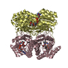
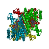
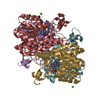
Deposited unit
A: Sorbitol-6-phosphate dehydrogenase
B: Sorbitol-6-phosphate dehydrogenase
C: Sorbitol-6-phosphate dehydrogenase
D: Sorbitol-6-phosphate dehydrogenase
E: Sorbitol-6-phosphate dehydrogenase
F: Sorbitol-6-phosphate dehydrogenase
G: Sorbitol-6-phosphate dehydrogenase
H: Sorbitol-6-phosphate dehydrogenase
I: Sorbitol-6-phosphate dehydrogenase
J: Sorbitol-6-phosphate dehydrogenase
K: Sorbitol-6-phosphate dehydrogenase
L: Sorbitol-6-phosphate dehydrogenase
hetero molecules Summary Component details
Theoretical mass Number of molelcules Total (without water) 338,370 24 Polymers 337,945 12 Non-polymers 425 12 Water 14,016 778
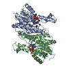
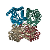
1
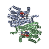
A: Sorbitol-6-phosphate dehydrogenase
B: Sorbitol-6-phosphate dehydrogenase
C: Sorbitol-6-phosphate dehydrogenase
D: Sorbitol-6-phosphate dehydrogenase
hetero molecules Summary Component details Symmetry operations Calculated values
Theoretical mass Number of molelcules Total (without water) 112,790 8 Polymers 112,648 4 Non-polymers 142 4 Water 72 4
Type Name Symmetry operation Number identity operation 1_555 x,y,z 1
Buried area 12980 Å2 ΔGint -153 kcal/mol Surface area 36420 Å2 Method
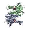
2
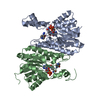
E: Sorbitol-6-phosphate dehydrogenase
F: Sorbitol-6-phosphate dehydrogenase
G: Sorbitol-6-phosphate dehydrogenase
H: Sorbitol-6-phosphate dehydrogenase
hetero molecules Summary Component details Symmetry operations Calculated values
Theoretical mass Number of molelcules Total (without water) 112,790 8 Polymers 112,648 4 Non-polymers 142 4 Water 72 4
Type Name Symmetry operation Number identity operation 1_555 x,y,z 1
Buried area 12950 Å2 ΔGint -156 kcal/mol Surface area 36120 Å2 Method
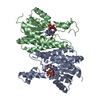
3
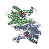
I: Sorbitol-6-phosphate dehydrogenase
J: Sorbitol-6-phosphate dehydrogenase
K: Sorbitol-6-phosphate dehydrogenase
L: Sorbitol-6-phosphate dehydrogenase
hetero molecules Summary Component details Symmetry operations Calculated values
Theoretical mass Number of molelcules Total (without water) 112,790 8 Polymers 112,648 4 Non-polymers 142 4 Water 72 4
Type Name Symmetry operation Number identity operation 1_555 x,y,z 1
Buried area 13020 Å2 ΔGint -152 kcal/mol Surface area 36340 Å2 Method
Unit cell Length a, b, c (Å) 120.707, 138.057, 199.442 Angle α, β, γ (deg.) 90.00, 90.00, 90.00 Int Tables number 19 Space group name H-M P21 21 21
 Movie
Movie Controller

 Yorodumi
 Open data
 Basic information
 Components
 Keywords
 Function and homology information
 Erwinia amylovora CFBP1430 (bacteria)
 X-RAY DIFFRACTION /
  Authors
 Italy, 1items
  Citation
 Structure visualization
 Downloads & links
 Other downloads

 PDBj
PDBj


 Assembly
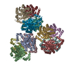


 Mass: 35.453 Da / Num. of mol.: 12 / Source method: obtained synthetically / Formula: Cl
 Mass: 18.015 Da / Num. of mol.: 778 / Source method: isolated from a natural source / Formula: H2O
 Sample preparation
 / Beamline: I04-1 / Wavelength: 0.9173 Å
 Processing