 Movie
Movie Controller

[English] 日本語
 Yorodumi
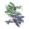
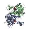
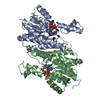
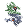
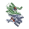
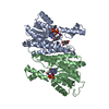
- PDB-1xr3: Actinorhodin Polyketide Ketoreductase with NADP and the Inhibitor... -
+ Open data

- Basic information
| Entry | Database: PDB / ID: 1xr3 | ||||||
|---|---|---|---|---|---|---|---|
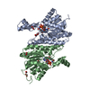
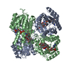
| Title | Actinorhodin Polyketide Ketoreductase with NADP and the Inhibitor Isoniazid bound | ||||||
 Components Components | ACTINORHODIN POLYKETIDE KETOREDUCTASE | ||||||
 Keywords Keywords | OXIDOREDUCTASE / PROTEIN-INHIBITOR COMPLEX | ||||||
| Function / homology |  Function and homology information Function and homology informationOxidoreductases; Acting on the CH-CH group of donors; With NAD+ or NADP+ as acceptor / monocarboxylic acid metabolic process / steroid metabolic process / antibiotic biosynthetic process / oxidoreductase activity Similarity search - Function | ||||||
| Biological species |  Streptomyces coelicolor (bacteria) Streptomyces coelicolor (bacteria) | ||||||
| Method |  X-RAY DIFFRACTION / SYNCHROTRON / MOLECULAR REPLACEMENT / Resolution: 2.71 Å X-RAY DIFFRACTION / SYNCHROTRON / MOLECULAR REPLACEMENT / Resolution: 2.71 Å | ||||||
 Authors Authors | Korman, T.P. / Hill, J.A. / Vu, T.N. / Tsai, S.C. | ||||||
 Citation Citation | Journal: Biochemistry / Year: 2004 Title: Structural analysis of actinorhodin polyketide ketoreductase: cofactor binding and substrate specificity. Authors: Korman, T.P. / Hill, J.A. / Vu, T.N. / Tsai, S.C. | ||||||
| History |
|
- Structure visualization
| Structure viewer | Molecule: MolmilJmol/JSmol |
|---|
- Downloads & links
-Download
| PDBx/mmCIF format | 1xr3.cif.gz | 106.9 KB | Display | PDBx/mmCIF format |
|---|---|---|---|---|
| PDB format | pdb1xr3.ent.gz | 82.6 KB | Display | PDB format |
| PDBx/mmJSON format | 1xr3.json.gz | Tree view | PDBx/mmJSON format | |
| Others |  Other downloads Other downloads |
-Validation report
| Arichive directory | https://data.pdbj.org/pub/pdb/validation_reports/xr/1xr3ftp://data.pdbj.org/pub/pdb/validation_reports/xr/1xr3 | HTTPS FTP |
|---|
-Related structure data
| Related structure data |  1x7gSC 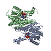 1x7hC S: Starting model for refinement C: citing same article ( |
|---|---|
| Similar structure data |

-Links
 PDBj
PDBj


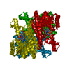
- Assembly
| Deposited unit | 
| ||||||||
|---|---|---|---|---|---|---|---|---|---|
| 1 | 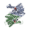
| ||||||||
| Unit cell |
| ||||||||
| Details | The biological assembly is a tetramer generated from the dimer in the asymmetric unit by the operations: -X, Y-X, 2/3-Z |
-Components
| #1: Protein | Mass: 27293.014 Da / Num. of mol.: 2 Source method: isolated from a genetically manipulated source Source: (gene. exp.) Streptomyces coelicolor (bacteria) / Gene: actIII / Plasmid: pET28c / Species (production host): Escherichia coli / Production host: References: UniProt: P16544, Oxidoreductases; Acting on the CH-CH group of donors; With NAD+ or NADP+ as acceptor #2: Chemical |   Mass: 743.405 Da / Num. of mol.: 2 / Source method: obtained synthetically / Formula: C21H28N7O17P3 Mass: 743.405 Da / Num. of mol.: 2 / Source method: obtained synthetically / Formula: C21H28N7O17P3#3: Chemical | 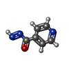  Mass: 135.123 Da / Num. of mol.: 2 / Source method: obtained synthetically / Formula: C6H5N3O / Comment: antibiotic*YM Mass: 135.123 Da / Num. of mol.: 2 / Source method: obtained synthetically / Formula: C6H5N3O / Comment: antibiotic*YM |
|---|
-Experimental details
-Experiment
| Experiment | Method: X-RAY DIFFRACTION / Number of used crystals: 2 |
|---|
- Sample preparation
| Crystal | Density Matthews: 3.43 Å3/Da / Density % sol: 64.14 % |
|---|---|
| Crystal grow | Temperature: 298 K / Method: vapor diffusion, sitting drop / pH: 7.5 Details: sodium formate, pH 7.5, VAPOR DIFFUSION, SITTING DROP, temperature 298K |
-Data collection
| Diffraction | Mean temperature: 75 K |
|---|---|
| Diffraction source | Source: SYNCHROTRON / Site: ALS  / Beamline: 5.0.1 / Wavelength: 1 Å / Beamline: 5.0.1 / Wavelength: 1 Å |
| Detector | Type: ADSC QUANTUM 4 / Detector: CCD / Date: Sep 11, 2004 / Details: mirrors |
| Radiation | Monochromator: Single crystal, cylindrically bent, Si(220) / Protocol: SINGLE WAVELENGTH / Monochromatic (M) / Laue (L): M / Scattering type: x-ray |
| Radiation wavelength | Wavelength: 1 Å / Relative weight: 1 |
| Reflection | Resolution: 2.71→50 Å / Num. obs: 21721 / % possible obs: 94.6 % / Observed criterion σ(I): -3 / Redundancy: 16.8 % / Biso Wilson estimate: 37.7 Å2 / Net I/σ(I): 16.1 |
| Reflection shell | Resolution: 2.71→2.76 Å / Redundancy: 7.2 % |
- Processing
| Software |
| |||||||||||||||||||||||||
|---|---|---|---|---|---|---|---|---|---|---|---|---|---|---|---|---|---|---|---|---|---|---|---|---|---|---|
| Refinement | Method to determine structure: MOLECULAR REPLACEMENT Starting model: PDB ENTRY 1X7G Resolution: 2.71→48.06 Å / Rfactor Rfree error: 0.006 / Data cutoff high absF: 74691.91 / Data cutoff low absF: 0 / Isotropic thermal model: RESTRAINED / Cross valid method: THROUGHOUT / σ(F): 0 / Stereochemistry target values: Engh & Huber
| |||||||||||||||||||||||||
| Solvent computation | Solvent model: FLAT MODEL / Bsol: 36.4248 Å2 / ksol: 0.359503 e/Å3 | |||||||||||||||||||||||||
| Displacement parameters | Biso mean: 41.3 Å2
| |||||||||||||||||||||||||
| Refine analyze |
| |||||||||||||||||||||||||
| Refinement step | Cycle: LAST / Resolution: 2.71→48.06 Å
| |||||||||||||||||||||||||
| Refine LS restraints |
| |||||||||||||||||||||||||
| LS refinement shell | Resolution: 2.71→2.88 Å / Rfactor Rfree error: 0.021 / Total num. of bins used: 6
| |||||||||||||||||||||||||
| Xplor file |
|
