| Entry | Database: PDB / ID: 5o2o
|
|---|
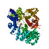
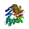
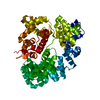
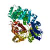
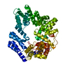
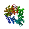
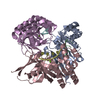
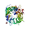
| Title | Lytic transglycosylase in action |
|---|
 Components Components | Transglycosylase |
|---|
 Keywords Keywords | HYDROLASE / lytic transglycosylases / acid/base catalysis / peptidoglycan / bacteria |
|---|
| Function / homology |  Function and homology information Function and homology information
catalytic activity / hydrolase activity, hydrolyzing O-glycosyl compounds / periplasmic space / metal ion bindingSimilarity search - Function Lytic transglycosylase, superhelical linker / Soluble lytic murein transglycosylase L domain / Soluble lytic transglycosylase helical domain / Lytic transglycosylase, superhelical U-shaped / Lytic transglycosylase, superhelical linker domain superfamily / Transglycosylase SLT domain 1 / Transglycosylase SLT domain / Lysozyme-like domain superfamily / Prokaryotic membrane lipoprotein lipid attachment site profile.Similarity search - Domain/homology |
|---|
| Biological species |  Neisseria meningitidis (bacteria) Neisseria meningitidis (bacteria) |
|---|
| Method |  X-RAY DIFFRACTION / SYNCHROTRON / MOLECULAR REPLACEMENT / Resolution: 1.43 Å X-RAY DIFFRACTION / SYNCHROTRON / MOLECULAR REPLACEMENT / Resolution: 1.43 Å |
|---|
 Authors Authors | Williams, A.H. / Hoauz, A. / Boneca, I.G. |
|---|
 Citation Citation | Journal: To Be Published
Title: Lytic transglycosylase in action
Authors: Williams, A.H. / Wheeler, R. / Rateau, L. / Malosse, C. / Rooke, J.C. / Hoauz, A. / Taha, M.-K. / Boneca, I.G. |
|---|
| History | | Deposition | May 22, 2017 | Deposition site: PDBE / Processing site: PDBE |
|---|
| Revision 1.0 | Jun 13, 2018 | Provider: repository / Type: Initial release |
|---|
| Revision 1.1 | Nov 28, 2018 | Group: Data collection / Structure summary / Category: audit_author |
|---|
| Revision 1.2 | Oct 16, 2019 | Group: Data collection / Category: reflns_shell |
|---|
| Revision 2.0 | Jul 29, 2020 | Group: Atomic model / Data collection ...Atomic model / Data collection / Derived calculations / Structure summary
Category: atom_site / chem_comp ...atom_site / chem_comp / entity / pdbx_branch_scheme / pdbx_chem_comp_identifier / pdbx_entity_branch / pdbx_entity_branch_descriptor / pdbx_entity_branch_link / pdbx_entity_branch_list / pdbx_entity_nonpoly / pdbx_nonpoly_scheme / pdbx_struct_assembly_gen / pdbx_struct_mod_residue / struct_asym / struct_conn / struct_site / struct_site_gen
Item: _atom_site.auth_asym_id / _atom_site.auth_seq_id ..._atom_site.auth_asym_id / _atom_site.auth_seq_id / _atom_site.label_asym_id / _chem_comp.name / _chem_comp.type / _entity.formula_weight / _entity.pdbx_description / _entity.pdbx_number_of_molecules / _entity.type / _pdbx_struct_assembly_gen.asym_id_list / _pdbx_struct_mod_residue.auth_asym_id / _pdbx_struct_mod_residue.auth_seq_id / _pdbx_struct_mod_residue.label_asym_id / _struct_conn.ptnr1_auth_asym_id / _struct_conn.ptnr1_auth_seq_id / _struct_conn.ptnr2_auth_asym_id / _struct_conn.ptnr2_auth_seq_id / _struct_conn.ptnr2_label_asym_id
Description: Carbohydrate remediation / Provider: repository / Type: Remediation |
|---|
| Revision 2.1 | Jan 17, 2024 | Group: Data collection / Database references ...Data collection / Database references / Refinement description / Structure summary
Category: chem_comp / chem_comp_atom ...chem_comp / chem_comp_atom / chem_comp_bond / database_2 / pdbx_initial_refinement_model
Item: _chem_comp.pdbx_synonyms / _database_2.pdbx_DOI / _database_2.pdbx_database_accession |
|---|
| Revision 2.2 | Oct 9, 2024 | Group: Structure summary / Category: pdbx_entry_details / pdbx_modification_feature |
|---|
|
|---|
 Movie
Movie Controller


 Open data
 Basic information
 Structure visualization
 Downloads & links
 Other downloads
 PDBj
PDBj


 Assembly
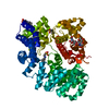

 Mass: 207.290 Da / Num. of mol.: 3 / Source method: obtained synthetically / Formula: C8H17NO3S / Comment: pH buffer*YM
 Mass: 18.015 Da / Num. of mol.: 847 / Source method: isolated from a natural source / Formula: H2O
 Sample preparation
 / Beamline: PROXIMA 1 / Wavelength: 1.1 Å
 Processing