 Movie
Movie Controller

[English] 日本語
 Yorodumi
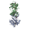
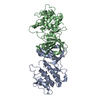
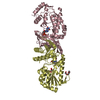
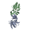
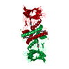
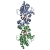
- PDB-5nx2: Crystal structure of thermostabilised full-length GLP-1R in compl... -
+ Open data

- Basic information
| Entry | Database: PDB / ID: 5nx2 | ||||||||||||
|---|---|---|---|---|---|---|---|---|---|---|---|---|---|
| Title | Crystal structure of thermostabilised full-length GLP-1R in complex with a truncated peptide agonist at 3.7 A resolution | ||||||||||||
 Components Components |
| ||||||||||||
 Keywords Keywords | MEMBRANE PROTEIN / 7TM / GPCR / signalling protein | ||||||||||||
| Function / homology |  Function and homology information Function and homology informationglucagon-like peptide 1 receptor activity / glucagon receptor activity / positive regulation of blood pressure / hormone secretion / post-translational protein targeting to membrane, translocation / response to psychosocial stress / regulation of heart contraction / peptide hormone binding / activation of adenylate cyclase activity / negative regulation of blood pressure ...glucagon-like peptide 1 receptor activity / glucagon receptor activity / positive regulation of blood pressure / hormone secretion / post-translational protein targeting to membrane, translocation / response to psychosocial stress / regulation of heart contraction / peptide hormone binding / activation of adenylate cyclase activity / negative regulation of blood pressure / Glucagon-type ligand receptors / transmembrane signaling receptor activity / Glucagon-like Peptide-1 (GLP1) regulates insulin secretion / adenylate cyclase-activating G protein-coupled receptor signaling pathway / positive regulation of cytosolic calcium ion concentration / G alpha (s) signalling events / learning or memory / cell surface receptor signaling pathway / membrane / plasma membrane Similarity search - Function | ||||||||||||
| Biological species |  Homo sapiens (human) Homo sapiens (human)synthetic construct (others) | ||||||||||||
| Method |  X-RAY DIFFRACTION / SYNCHROTRON / MOLECULAR REPLACEMENT / Resolution: 3.7 Å X-RAY DIFFRACTION / SYNCHROTRON / MOLECULAR REPLACEMENT / Resolution: 3.7 Å | ||||||||||||
 Authors Authors | Rappas, M. / Jazayeri, A. / Brown, A.J.H. / Kean, J. / Errey, J.C. / Robertson, N. / Fiez-Vandal, C. / Andrews, S.P. / Congreve, M. / Bortolato, A. ...Rappas, M. / Jazayeri, A. / Brown, A.J.H. / Kean, J. / Errey, J.C. / Robertson, N. / Fiez-Vandal, C. / Andrews, S.P. / Congreve, M. / Bortolato, A. / Mason, J.S. / Baig, A.H. / Teobald, I. / Dore, A.S. / Weir, M. / Cooke, R.M. / Marshall, F.H. | ||||||||||||
 Citation Citation | Journal: Nature / Year: 2017 Title: Crystal structure of the GLP-1 receptor bound to a peptide agonist. Authors: Jazayeri, A. / Rappas, M. / Brown, A.J.H. / Kean, J. / Errey, J.C. / Robertson, N.J. / Fiez-Vandal, C. / Andrews, S.P. / Congreve, M. / Bortolato, A. / Mason, J.S. / Baig, A.H. / Teobald, I. ...Authors: Jazayeri, A. / Rappas, M. / Brown, A.J.H. / Kean, J. / Errey, J.C. / Robertson, N.J. / Fiez-Vandal, C. / Andrews, S.P. / Congreve, M. / Bortolato, A. / Mason, J.S. / Baig, A.H. / Teobald, I. / Dore, A.S. / Weir, M. / Cooke, R.M. / Marshall, F.H. | ||||||||||||
| History |
|
- Structure visualization
| Structure viewer | Molecule: MolmilJmol/JSmol |
|---|
- Downloads & links
-Download
| PDBx/mmCIF format | 5nx2.cif.gz | 190.8 KB | Display | PDBx/mmCIF format |
|---|---|---|---|---|
| PDB format | pdb5nx2.ent.gz | 150.7 KB | Display | PDB format |
| PDBx/mmJSON format | 5nx2.json.gz | Tree view | PDBx/mmJSON format | |
| Others |  Other downloads Other downloads |
-Validation report
| Arichive directory | https://data.pdbj.org/pub/pdb/validation_reports/nx/5nx2ftp://data.pdbj.org/pub/pdb/validation_reports/nx/5nx2 | HTTPS FTP |
|---|
-Related structure data


-Links
 PDBj
PDBj


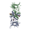
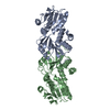
- Assembly
| Deposited unit | 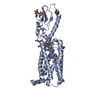
| ||||||||
|---|---|---|---|---|---|---|---|---|---|
| 1 |
| ||||||||
| Unit cell |
|
-Components
| #1: Protein | Mass: 49028.711 Da / Num. of mol.: 1 / Fragment: UNP residues 24-432 Mutation: T207E, Q211A, D215R, L232F, G295A, T298A, C329A, P358A, G361A, H363V, V405A Source method: isolated from a genetically manipulated source Source: (gene. exp.) Homo sapiens (human) / Gene: GLP1R / Plasmid: pFastBac1-HM / Production host:   Spodoptera frugiperda (fall armyworm) / References: UniProt: P43220 Spodoptera frugiperda (fall armyworm) / References: UniProt: P43220 |
|---|---|
| #2: Protein/peptide | Mass: 1488.620 Da / Num. of mol.: 1 / Source method: obtained synthetically / Source: (synth.) synthetic construct (others) |
| #3: Polysaccharide | 2-acetamido-2-deoxy-beta-D-glucopyranose-(1-4)-2-acetamido-2-deoxy-beta-D-glucopyranose Source method: isolated from a genetically manipulated source |
| #4: Sugar | ChemComp-NAG /   Type: D-saccharide, beta linking / Mass: 221.208 Da / Num. of mol.: 1 Type: D-saccharide, beta linking / Mass: 221.208 Da / Num. of mol.: 1Source method: isolated from a genetically manipulated source Formula: C8H15NO6 |
| #5: Sugar | 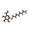  Type: D-saccharide / Mass: 308.434 Da / Num. of mol.: 3 Type: D-saccharide / Mass: 308.434 Da / Num. of mol.: 3Source method: isolated from a genetically manipulated source Formula: C14H28O5S / Comment: detergent*YM |
-Experimental details
-Experiment
| Experiment | Method: X-RAY DIFFRACTION / Number of used crystals: 1 |
|---|
- Sample preparation
| Crystal | Density Matthews: 4.31 Å3/Da / Density % sol: 71.46 % / Description: Rod-like crystals |
|---|---|
| Crystal grow | Temperature: 283 K / Method: vapor diffusion, sitting drop / Details: 0.1M Tris-HCl pH 8.0-9.0, 32-44% (v/v) PEG 200 / PH range: 8.0 - 9.0 |
-Data collection
| Diffraction | Mean temperature: 100 K / Ambient temp details: Liquid nitrogen cryojet |
|---|---|
| Diffraction source | Source: SYNCHROTRON / Site: Diamond  / Beamline: I24 / Wavelength: 0.96861 Å / Beamline: I24 / Wavelength: 0.96861 Å |
| Detector | Type: DECTRIS PILATUS3 6M / Detector: PIXEL / Date: Dec 11, 2015 Details: Oxford Danfysik/SESO Two stage demagnification using two K-B pairs of bimorph type mirrors |
| Radiation | Monochromator: ACCEL Fixed exit Double Crystal Si(111) / Protocol: SINGLE WAVELENGTH / Monochromatic (M) / Laue (L): M / Scattering type: x-ray |
| Radiation wavelength | Wavelength: 0.96861 Å / Relative weight: 1 |
| Reflection | Resolution: 3.7→54.6 Å / Num. obs: 9266 / % possible obs: 98.5 % / Redundancy: 6.6 % / Biso Wilson estimate: 135 Å2 / CC1/2: 0.998 / Rmerge(I) obs: 0.134 / Rpim(I) all: 0.073 / Net I/σ(I): 6.6 |
| Reflection shell | Resolution: 3.7→4.05 Å / Redundancy: 6.9 % / Rmerge(I) obs: 2.024 / Mean I/σ(I) obs: 1.2 / Num. unique obs: 2205 / CC1/2: 0.348 / Rpim(I) all: 1.078 / % possible all: 99.7 |
- Processing
| Software |
| ||||||||||||||||||||||||||||||||||||||||||||||||||||||||||||||||||||||||||||||||||||||||||||||||||||
|---|---|---|---|---|---|---|---|---|---|---|---|---|---|---|---|---|---|---|---|---|---|---|---|---|---|---|---|---|---|---|---|---|---|---|---|---|---|---|---|---|---|---|---|---|---|---|---|---|---|---|---|---|---|---|---|---|---|---|---|---|---|---|---|---|---|---|---|---|---|---|---|---|---|---|---|---|---|---|---|---|---|---|---|---|---|---|---|---|---|---|---|---|---|---|---|---|---|---|---|---|---|
| Refinement | Method to determine structure: MOLECULAR REPLACEMENT Starting model: 3IOL, 5EE7 Resolution: 3.7→24.677 Å / SU ML: 0.81 / Cross valid method: FREE R-VALUE / σ(F): 0.02 / Phase error: 46.76 / Stereochemistry target values: ML
| ||||||||||||||||||||||||||||||||||||||||||||||||||||||||||||||||||||||||||||||||||||||||||||||||||||
| Solvent computation | Shrinkage radii: 0.9 Å / VDW probe radii: 1.11 Å / Solvent model: FLAT BULK SOLVENT MODEL | ||||||||||||||||||||||||||||||||||||||||||||||||||||||||||||||||||||||||||||||||||||||||||||||||||||
| Refinement step | Cycle: LAST / Resolution: 3.7→24.677 Å
| ||||||||||||||||||||||||||||||||||||||||||||||||||||||||||||||||||||||||||||||||||||||||||||||||||||
| Refine LS restraints |
| ||||||||||||||||||||||||||||||||||||||||||||||||||||||||||||||||||||||||||||||||||||||||||||||||||||
| LS refinement shell |
| ||||||||||||||||||||||||||||||||||||||||||||||||||||||||||||||||||||||||||||||||||||||||||||||||||||
| Refinement TLS params. | Method: refined / Refine-ID: X-RAY DIFFRACTION
| ||||||||||||||||||||||||||||||||||||||||||||||||||||||||||||||||||||||||||||||||||||||||||||||||||||
| Refinement TLS group |
|
