Mass: 18.015 Da / Num. of mol.: 310 / Source method: isolated from a natural source / Formula: H2O
Has protein modification
Y
-
Experimental details
-
Experiment
Experiment
Method: X-RAY DIFFRACTION / Number of used crystals: 1
-
Sample preparation
Crystal
Density Matthews: 2.34 Å3/Da / Density % sol: 47 %
Crystal grow
Temperature: 294.15 K / Method: microbatch / pH: 5.5 Details: 2 micro liters of 18 mg/ml protein solution containing 1 mM XMP, 1 mM PPi, 20 mM MgCl2 was mixed with an equal volume of crystallization condition containing 20 % (w/v) PEG 1500 and 0.1 M MIB buffer pH 5.5
-
Data collection
Diffraction
Mean temperature: 100 K / Serial crystal experiment: N
Diffraction source
Source: ROTATING ANODE / Type: BRUKER AXS MICROSTAR / Wavelength: 1.54 Å
Detector
Type: MAR scanner 345 mm plate / Detector: IMAGE PLATE / Date: Oct 23, 2017
Radiation
Protocol: SINGLE WAVELENGTH / Monochromatic (M) / Laue (L): M / Scattering type: x-ray
Radiation wavelength
Wavelength: 1.54 Å / Relative weight: 1
Reflection
Resolution: 2.1→52.53 Å / Num. obs: 76858 / % possible obs: 100 % / Redundancy: 7.6 % / Biso Wilson estimate: 32.1 Å2 / CC1/2: 0.989 / Net I/σ(I): 8.9
Reflection shell
Resolution: 2.1→2.14 Å / Redundancy: 7.5 % / Mean I/σ(I) obs: 1.7 / Num. unique obs: 4500 / CC1/2: 0.645 / % possible all: 100
-
Processing
Software
Name
Version
Classification
REFMAC
5.8.0158
refinement
iMOSFLM
datareduction
Aimless
datascaling
PHASER
phasing
Refinement
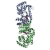
Method to determine structure: MOLECULAR REPLACEMENT Starting model: 2DPL
 Movie
Movie Controller

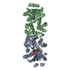
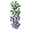
 Yorodumi
 Open data
 Basic information
 Components
 Keywords
 Function and homology information
 Methanocaldococcus jannaschii DSM 2661 (archaea)
 X-RAY DIFFRACTION /
  Authors
 India, 2items
  Citation
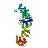
 Structure visualization
 Downloads & links
 Other downloads


 PDBj
PDBj
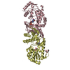
 Assembly
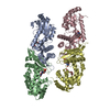

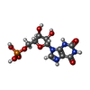
 Mass: 365.213 Da / Num. of mol.: 4 / Source method: obtained synthetically / Formula: C10H14N4O9P / Feature type: SUBJECT OF INVESTIGATION
 Mass: 150.173 Da / Num. of mol.: 1 / Source method: obtained synthetically / Formula: C6H14O4
 Mass: 104.061 Da / Num. of mol.: 1 / Source method: obtained synthetically / Formula: C3H4O4
 Mass: 18.015 Da / Num. of mol.: 310 / Source method: isolated from a natural source / Formula: H2O
 Sample preparation
 Processing