[English] 日本語
 Yorodumi
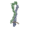
- PDB-5nw5: Crystal structure of the Rif1 N-terminal domain (RIF1-NTD) from S... -
+ Open data
- Basic information
| Entry | Database: PDB / ID: 5nw5 | ||||||
|---|---|---|---|---|---|---|---|
| Title | Crystal structure of the Rif1 N-terminal domain (RIF1-NTD) from Saccharomyces cerevisiae in complex with DNA | ||||||
 Components Components |
| ||||||
 Keywords Keywords | DNA BINDING PROTEIN / telomere maintenance / DNA double-strand break repair / irregular helical repeat / all-alpha fold | ||||||
| Function / homology |  Function and homology information Function and homology informationnegative regulation of mitotic DNA replication initiation from late origin / regulation of DNA stability / chromosome, telomeric repeat region / shelterin complex / centromeric DNA binding / DNA double-strand break processing / telomere capping / silent mating-type cassette heterochromatin formation / protein localization to chromosome, telomeric region / telomeric DNA binding ...negative regulation of mitotic DNA replication initiation from late origin / regulation of DNA stability / chromosome, telomeric repeat region / shelterin complex / centromeric DNA binding / DNA double-strand break processing / telomere capping / silent mating-type cassette heterochromatin formation / protein localization to chromosome, telomeric region / telomeric DNA binding / DNA replication origin binding / DNA replication initiation / RNA polymerase II core promoter sequence-specific DNA binding / negative regulation of DNA-templated DNA replication initiation / telomere maintenance / chromosome, telomeric region / nucleus Similarity search - Function | ||||||
| Biological species |  synthetic construct (others) | ||||||
| Method |  X-RAY DIFFRACTION / X-RAY DIFFRACTION /  SYNCHROTRON / SYNCHROTRON /  MOLECULAR REPLACEMENT / Resolution: 6.502 Å MOLECULAR REPLACEMENT / Resolution: 6.502 Å | ||||||
 Authors Authors | Bunker, R.D. / Reinert, J.K. / Shi, T. / Thoma, N.H. | ||||||
 Citation Citation |  Journal: Nat. Struct. Mol. Biol. / Year: 2017 Journal: Nat. Struct. Mol. Biol. / Year: 2017Title: Rif1 maintains telomeres and mediates DNA repair by encasing DNA ends. Authors: Mattarocci, S. / Reinert, J.K. / Bunker, R.D. / Fontana, G.A. / Shi, T. / Klein, D. / Cavadini, S. / Faty, M. / Shyian, M. / Hafner, L. / Shore, D. / Thoma, N.H. / Rass, U. | ||||||
| History |
|
- Structure visualization
| Structure viewer | Molecule:  Molmil Molmil Jmol/JSmol Jmol/JSmol |
|---|
- Downloads & links
- Download
| PDBx/mmCIF format |  5nw5.cif.gz 5nw5.cif.gz | 795 KB | Display |  PDBx/mmCIF format PDBx/mmCIF format |
|---|---|---|---|---|
| PDB format |  pdb5nw5.ent.gz pdb5nw5.ent.gz | 629.8 KB | Display |  PDB format PDB format |
| PDBx/mmJSON format |  5nw5.json.gz 5nw5.json.gz | Tree view |  PDBx/mmJSON format PDBx/mmJSON format | |
| Others |  Other downloads Other downloads |
-Validation report
| Summary document |  5nw5_validation.pdf.gz 5nw5_validation.pdf.gz | 436 KB | Display |  wwPDB validaton report wwPDB validaton report |
|---|---|---|---|---|
| Full document |  5nw5_full_validation.pdf.gz 5nw5_full_validation.pdf.gz | 475.1 KB | Display | |
| Data in XML |  5nw5_validation.xml.gz 5nw5_validation.xml.gz | 41.2 KB | Display | |
| Data in CIF |  5nw5_validation.cif.gz 5nw5_validation.cif.gz | 63.7 KB | Display | |
| Arichive directory |  https://data.pdbj.org/pub/pdb/validation_reports/nw/5nw5 https://data.pdbj.org/pub/pdb/validation_reports/nw/5nw5 ftp://data.pdbj.org/pub/pdb/validation_reports/nw/5nw5 ftp://data.pdbj.org/pub/pdb/validation_reports/nw/5nw5 | HTTPS FTP |
-Related structure data
| Related structure data |  5nvrSC S: Starting model for refinement C: citing same article ( |
|---|---|
| Similar structure data | |
| Experimental dataset #1 | Data reference:  10.15785/SBGRID/446 / Data set type: diffraction image data 10.15785/SBGRID/446 / Data set type: diffraction image data |
- Links
Links
- Assembly
| Deposited unit | 
| ||||||||
|---|---|---|---|---|---|---|---|---|---|
| 1 |
| ||||||||
| Unit cell |
|
- Components
| #1: Protein | Mass: 140781.562 Da / Num. of mol.: 2 Source method: isolated from a genetically manipulated source Source: (gene. exp.)   Trichoplusia ni (cabbage looper) / References: UniProt: P29539 Trichoplusia ni (cabbage looper) / References: UniProt: P29539#2: DNA chain | | Mass: 9351.247 Da / Num. of mol.: 1 / Source method: obtained synthetically Details: Directionality and sequence register of the DNA could not be established unequivocally. DNA duplex modeled as poly-T in the most plausible orientation. Chain C construct sequence: ...Details: Directionality and sequence register of the DNA could not be established unequivocally. DNA duplex modeled as poly-T in the most plausible orientation. Chain C construct sequence: ACGCTGCCGAATTCTACCAGTGCCTTGCTAGGACATCTTTGCCCACCTGCAGGTTCACCC. Source: (synth.) synthetic construct (others) #3: DNA chain | | Mass: 9080.827 Da / Num. of mol.: 1 / Source method: obtained synthetically Details: Directionality and sequence register of the DNA could not be established unequivocally. DNA duplex modeled as poly-T in most plausible orientation. Chain D construct sequence: TAGCAAGGCACTGGTAGAATTCGGCAGCGT. Source: (synth.) synthetic construct (others) |
|---|
-Experimental details
-Experiment
| Experiment | Method:  X-RAY DIFFRACTION / Number of used crystals: 1 X-RAY DIFFRACTION / Number of used crystals: 1 |
|---|
- Sample preparation
| Crystal | Density Matthews: 5.09 Å3/Da / Density % sol: 75.82 % |
|---|---|
| Crystal grow | Temperature: 295 K / Method: vapor diffusion, sitting drop / pH: 7.4 Details: CRYSTAL GROWN BY DEHYDRATING 1 uL of PROTEIN-DNA MIXTURE (43.5 uM protein with 65 uM DNA) IN 50 mM HEPES PH 7.4, 310 mM NaCl, 1 mM TCEP over a reservoir containing 10 mM NiCl2, 100 mM Tris- ...Details: CRYSTAL GROWN BY DEHYDRATING 1 uL of PROTEIN-DNA MIXTURE (43.5 uM protein with 65 uM DNA) IN 50 mM HEPES PH 7.4, 310 mM NaCl, 1 mM TCEP over a reservoir containing 10 mM NiCl2, 100 mM Tris-HCl, pH 8, 20% (w/v) polyethylene glycol MME 2000 |
-Data collection
| Diffraction | Mean temperature: 100 K |
|---|---|
| Diffraction source | Source:  SYNCHROTRON / Site: SYNCHROTRON / Site:  SLS SLS  / Beamline: X06DA / Wavelength: 0.91863 Å / Beamline: X06DA / Wavelength: 0.91863 Å |
| Detector | Type: DECTRIS PILATUS 2M-F / Detector: PIXEL / Date: Feb 26, 2013 / Details: DYNAMICALLY BENDABLE MIRROR |
| Radiation | Monochromator: SI(111) / Protocol: SINGLE WAVELENGTH / Monochromatic (M) / Laue (L): M / Scattering type: x-ray |
| Radiation wavelength | Wavelength: 0.91863 Å / Relative weight: 1 |
| Reflection | Resolution: 6.5→43.25 Å / Num. obs: 12703 / % possible obs: 99.6 % / Observed criterion σ(I): -3 / Redundancy: 11.9 % / Biso Wilson estimate: 388 Å2 / CC1/2: 0.998 / Rmerge(I) obs: 0.342 / Rpim(I) all: 0.102 / Net I/σ(I): 8 |
| Reflection shell | Resolution: 6.5→7.27 Å / Redundancy: 12.5 % / Rmerge(I) obs: 6.011 / Mean I/σ(I) obs: 0.5 / Num. unique obs: 3529 / CC1/2: 0.142 / Rpim(I) all: 1.757 / % possible all: 100 |
- Processing
| Software |
| |||||||||||||||||||||||||||||||||||
|---|---|---|---|---|---|---|---|---|---|---|---|---|---|---|---|---|---|---|---|---|---|---|---|---|---|---|---|---|---|---|---|---|---|---|---|---|
| Refinement | Method to determine structure:  MOLECULAR REPLACEMENT MOLECULAR REPLACEMENTStarting model: 5NVR Resolution: 6.502→43.232 Å / SU ML: 1.11 / Cross valid method: THROUGHOUT / σ(F): 1.35 / Phase error: 32.41 Details: ANISOTROPICALLY TRUNCATED STRUCTURE FACTOR AMPLITUDES GENERATED BY STARANISO USED FOR REFINEMENT. FINAL REFINEMENT CARRIED OUT WITH HYBRID PHENIX/AMBER.
| |||||||||||||||||||||||||||||||||||
| Solvent computation | Shrinkage radii: 0.9 Å / VDW probe radii: 1.11 Å | |||||||||||||||||||||||||||||||||||
| Displacement parameters | Biso mean: 331 Å2 | |||||||||||||||||||||||||||||||||||
| Refinement step | Cycle: LAST / Resolution: 6.502→43.232 Å
| |||||||||||||||||||||||||||||||||||
| Refine LS restraints |
| |||||||||||||||||||||||||||||||||||
| LS refinement shell |
|
 Movie
Movie Controller


 PDBj
PDBj


