 Movie
Movie Controller


+ Open data

- Basic information
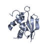
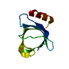
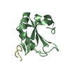
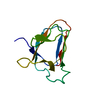
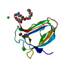
| Entry | Database: PDB / ID: 5ntb | ||||||
|---|---|---|---|---|---|---|---|
| Title | Streptomyces papain inhibitor (SPI) | ||||||
 Components Components | Papain inhibitor | ||||||
 Keywords Keywords | Protease inhibitor / Streptomyces papain inhibitor / transglutaminase / glutamine donor / Streptomyces mobaraensis | ||||||
| Function / homology |  Function and homology information Function and homology informationnegative regulation of peptidase activity / cysteine-type endopeptidase inhibitor activity / serine-type endopeptidase inhibitor activity / : Similarity search - Function | ||||||
| Biological species |  Streptomyces mobaraensis (bacteria) Streptomyces mobaraensis (bacteria) | ||||||
| Method |  X-RAY DIFFRACTION / SYNCHROTRON / MOLECULAR REPLACEMENT / Resolution: 1.5 Å X-RAY DIFFRACTION / SYNCHROTRON / MOLECULAR REPLACEMENT / Resolution: 1.5 Å | ||||||
 Authors Authors | Schmelz, S. / Juettner, N.E. / Fuchsbauer, H.L. / Scrima, A. | ||||||
| Funding support |  Germany, 1items Germany, 1items
| ||||||
 Citation Citation | Journal: Protein Sci. / Year: 2018 Title: Illuminating structure and acyl donor sites of a physiological transglutaminase substrate from Streptomyces mobaraensis. Authors: Juettner, N.E. / Schmelz, S. / Bogen, J.P. / Happel, D. / Fessner, W.D. / Pfeifer, F. / Fuchsbauer, H.L. / Scrima, A. | ||||||
| History |
|
- Structure visualization
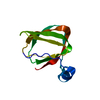
| Structure viewer | Molecule: MolmilJmol/JSmol |
|---|
- Downloads & links
-Download
| PDBx/mmCIF format | 5ntb.cif.gz | 61.8 KB | Display | PDBx/mmCIF format |
|---|---|---|---|---|
| PDB format | pdb5ntb.ent.gz | 44.1 KB | Display | PDB format |
| PDBx/mmJSON format | 5ntb.json.gz | Tree view | PDBx/mmJSON format | |
| Others |  Other downloads Other downloads |
-Validation report
| Arichive directory | https://data.pdbj.org/pub/pdb/validation_reports/nt/5ntbftp://data.pdbj.org/pub/pdb/validation_reports/nt/5ntb | HTTPS FTP |
|---|
-Related structure data
| Related structure data |  4jcwS S: Starting model for refinement |
|---|---|
| Similar structure data |


-Links
 PDBj
PDBj- Assembly
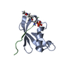
| Deposited unit | 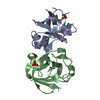
| ||||||||
|---|---|---|---|---|---|---|---|---|---|
| 1 | 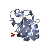
| ||||||||
| 2 | 
| ||||||||
| Unit cell |
|
-Components
| #1: Protein | Mass: 12041.707 Da / Num. of mol.: 2 / Source method: isolated from a natural source / Source: (natural) Streptomyces mobaraensis (bacteria) / References: UniProt: P86242#2: Chemical |   Mass: 96.063 Da / Num. of mol.: 3 / Source method: obtained synthetically / Formula: SO4 Mass: 96.063 Da / Num. of mol.: 3 / Source method: obtained synthetically / Formula: SO4#3: Water | ChemComp-HOH / |  Mass: 18.015 Da / Num. of mol.: 232 / Source method: isolated from a natural source / Formula: H2O Mass: 18.015 Da / Num. of mol.: 232 / Source method: isolated from a natural source / Formula: H2OHas protein modification | Y | |
|---|
-Experimental details
-Experiment
| Experiment | Method: X-RAY DIFFRACTION / Number of used crystals: 1 |
|---|
- Sample preparation
| Crystal | Density Matthews: 1.92 Å3/Da / Density % sol: 35.9 % |
|---|---|
| Crystal grow | Temperature: 292 K / Method: vapor diffusion, sitting drop / Details: 2.2M Ammonium sulphate, 200 mM Litium chloride |
-Data collection
| Diffraction | Mean temperature: 100 K |
|---|---|
| Diffraction source | Source: SYNCHROTRON / Site: ESRF  / Beamline: ID30B / Wavelength: 1.007 Å / Beamline: ID30B / Wavelength: 1.007 Å |
| Detector | Type: DECTRIS PILATUS 6M-F / Detector: PIXEL / Date: Dec 14, 2015 |
| Radiation | Protocol: SINGLE WAVELENGTH / Monochromatic (M) / Laue (L): M / Scattering type: x-ray |
| Radiation wavelength | Wavelength: 1.007 Å / Relative weight: 1 |
| Reflection | Resolution: 1.5→41.9 Å / Num. obs: 31262 / % possible obs: 99.8 % / Redundancy: 12.2 % / CC1/2: 0.999 / Rmerge(I) obs: 0.09 / Net I/σ(I): 16.6 |
| Reflection shell | Resolution: 1.5→1.6 Å / Redundancy: 12 % / Rmerge(I) obs: 1.03 / Mean I/σ(I) obs: 3.1 / Num. unique obs: 5375 / CC1/2: 0.789 / % possible all: 99.8 |
- Processing
| Software |
| ||||||||||||||||||||||||||||||||||||||||||||||||||||||||||||||||||||||||||||||||||||
|---|---|---|---|---|---|---|---|---|---|---|---|---|---|---|---|---|---|---|---|---|---|---|---|---|---|---|---|---|---|---|---|---|---|---|---|---|---|---|---|---|---|---|---|---|---|---|---|---|---|---|---|---|---|---|---|---|---|---|---|---|---|---|---|---|---|---|---|---|---|---|---|---|---|---|---|---|---|---|---|---|---|---|---|---|---|
| Refinement | Method to determine structure: MOLECULAR REPLACEMENT Starting model: 4JCW Resolution: 1.5→41.863 Å / SU ML: 0.13 / Cross valid method: FREE R-VALUE / σ(F): 1.35 / Phase error: 17.97
| ||||||||||||||||||||||||||||||||||||||||||||||||||||||||||||||||||||||||||||||||||||
| Solvent computation | Shrinkage radii: 0.9 Å / VDW probe radii: 1.11 Å | ||||||||||||||||||||||||||||||||||||||||||||||||||||||||||||||||||||||||||||||||||||
| Refinement step | Cycle: LAST / Resolution: 1.5→41.863 Å
| ||||||||||||||||||||||||||||||||||||||||||||||||||||||||||||||||||||||||||||||||||||
| Refine LS restraints |
| ||||||||||||||||||||||||||||||||||||||||||||||||||||||||||||||||||||||||||||||||||||
| LS refinement shell |
|
