[English] 日本語
Yorodumi
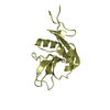
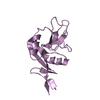
- PDB-5nsa: Beta domain of human transcobalamin bound to Co-beta-[2-(2,4-difl... -

+
Open data


ID or keywords:

Loading...

-
Basic information

Entry
Database: PDB / ID: 5nsa
TitleBeta domain of human transcobalamin bound to Co-beta-[2-(2,4-difluorophenyl)ethinyl]cobalamin
ComponentsTranscobalamin-2
KeywordsTRANSPORT PROTEIN / binding protein / bloodstream / antivitamin B12
Function / homology
Function and homology information


Defective TCN2 causes TCN2 deficiency / Defective CD320 causes MMATC / Transport of RCbl within the body / cargo receptor ligand activity / cobalt ion transport / cobalamin transport / cobalamin binding / lysosomal lumen / external side of plasma membrane / extracellular space ...Defective TCN2 causes TCN2 deficiency / Defective CD320 causes MMATC / Transport of RCbl within the body / cargo receptor ligand activity / cobalt ion transport / cobalamin transport / cobalamin binding / lysosomal lumen / external side of plasma membrane / extracellular space / extracellular region / metal ion binding / plasma membrane
Similarity search - Function
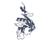
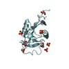
Domain of unknown function DUF4430 / Ferric Hydroxamate Uptake Protein; Chain A, domain 1 - #30 / Domain of unknown function (DUF4430) / Cobalamin (vitamin B12)-binding protein / Eukaryotic cobalamin-binding protein / Eukaryotic cobalamin-binding proteins signature. / Ferric Hydroxamate Uptake Protein; Chain A, domain 1 / Terpenoid cyclases/protein prenyltransferase alpha-alpha toroid / Beta Complex / Mainly Beta
Similarity search - Domain/homology
1-ethynyl-2,4-difluorobenzene / COBALAMIN / Transcobalamin-2
Similarity search - Component
Biological speciesHomo sapiens (human)
MethodX-RAY DIFFRACTION / SYNCHROTRON / MOLECULAR REPLACEMENT / Resolution: 1.273 Å
AuthorsBloch, J.S. / Ruetz, M. / Krautler, B. / Locher, K.P.
Funding support Switzerland, 1items
OrganizationGrant numberCountry
Swiss National Science Foundation310030B_166672 Switzerland
CitationJournal: PLoS ONE / Year: 2017
Title: Structure of the human transcobalamin beta domain in four distinct states.
Authors: Bloch, J.S. / Ruetz, M. / Krautler, B. / Locher, K.P.
History
DepositionApr 25, 2017Deposition site: PDBE / Processing site: PDBE
Revision 1.0Sep 27, 2017Provider: repository / Type: Initial release
Revision 1.1Oct 16, 2019Group: Data collection / Category: reflns_shell

-
Structure visualization

Structure viewerMolecule:
MolmilJmol/JSmol

Downloads & links

-
Assembly

Deposited unit
A: Transcobalamin-2
hetero molecules


Theoretical massNumber of molelcules
Total (without water)13,4784
Polymers11,9701
Non-polymers1,5093
Water4,252236
1


  • Idetical with deposited unit
  • defined by author&software
TypeNameSymmetry operationNumber
identity operation1_555x,y,z1
Buried area1200 Å2
ΔGint-17 kcal/mol
Surface area6980 Å2
MethodPISA
Unit cell
Length a, b, c (Å)61.721, 62.298, 33.762
Angle α, β, γ (deg.)90.00, 90.00, 90.00
Int Tables number18
Space group name H-MP21212
Components on special symmetry positions
IDModelComponents
11A-679-

HOH

-
Components

#1: Protein Transcobalamin-2 / / TC-2 / Transcobalamin II / TCII


Mass: 11969.556 Da / Num. of mol.: 1
Source method: isolated from a genetically manipulated source
Source: (gene. exp.) Homo sapiens (human) / Gene: TCN2, TC2 / Production host: 
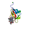
Spodoptera frugiperda (fall armyworm) / References: UniProt: P20062
#2: Chemical ChemComp-B12 / COBALAMIN / Vitamin B12


Mass: 1330.356 Da / Num. of mol.: 1 / Source method: obtained synthetically / Formula: C62H89CoN13O14P
#3: Chemical ChemComp-8FS / 1-ethynyl-2,4-difluorobenzene


Mass: 138.114 Da / Num. of mol.: 1 / Source method: obtained synthetically / Formula: C8H4F2
#4: Chemical ChemComp-CA / CALCIUM ION


Mass: 40.078 Da / Num. of mol.: 1 / Source method: obtained synthetically / Formula: Ca
#5: Water ChemComp-HOH / water / Water


Mass: 18.015 Da / Num. of mol.: 236 / Source method: isolated from a natural source / Formula: H2O

-
Experimental details

-
Experiment

ExperimentMethod: X-RAY DIFFRACTION / Number of used crystals: 1

-
Sample preparation

CrystalDensity Matthews: 2.71 Å3/Da / Density % sol: 54.64 %
Crystal growTemperature: 293.15 K / Method: evaporation
Details: 0.3 M potassium formate, 17% w/v polyethylene glycol 3,350

-
Data collection

DiffractionMean temperature: 100 K
Diffraction sourceSource: SYNCHROTRON / Site: SLS / Beamline: X10SA / Wavelength: 1 Å
DetectorType: PSI PILATUS 6M / Detector: PIXEL / Date: Sep 30, 2016
RadiationProtocol: SINGLE WAVELENGTH / Monochromatic (M) / Laue (L): M / Scattering type: x-ray
Radiation wavelengthWavelength: 1 Å / Relative weight: 1
ReflectionResolution: 1.27→43.85 Å / Num. obs: 33543 / % possible obs: 96.19 % / Redundancy: 6.3 % / Net I/σ(I): 24.4

-
Processing

Software
NameVersionClassification
PHENIX(1.12_2829: ???)refinement
XDSdata scaling
PHENIXphasing
RefinementMethod to determine structure: MOLECULAR REPLACEMENT / Resolution: 1.273→43.846 Å / SU ML: 0.17 / Cross valid method: FREE R-VALUE / σ(F): 1.33 / Phase error: 19.57 / Stereochemistry target values: ML
RfactorNum. reflection% reflection
Rfree0.1788 3112 4.98 %
Rwork0.1442 --
obs0.1459 62434 94.74 %
Solvent computationShrinkage radii: 0.9 Å / VDW probe radii: 1.11 Å / Solvent model: FLAT BULK SOLVENT MODEL
Refinement stepCycle: LAST / Resolution: 1.273→43.846 Å
ProteinNucleic acidLigandSolventTotal
Num. atoms827 0 102 236 1165
Refine LS restraints
Refine-IDTypeDev idealNumber
X-RAY DIFFRACTIONf_bond_d0.0161028
X-RAY DIFFRACTIONf_angle_d2.2841439
X-RAY DIFFRACTIONf_dihedral_angle_d20.713396
X-RAY DIFFRACTIONf_chiral_restr0.629160
X-RAY DIFFRACTIONf_plane_restr0.009184
LS refinement shell
Resolution (Å)Rfactor RfreeNum. reflection RfreeRfactor RworkNum. reflection RworkRefine-ID% reflection obs (%)
1.2733-1.29320.4031950.39931767X-RAY DIFFRACTION61
1.2932-1.31440.5625940.54541828X-RAY DIFFRACTION65
1.3144-1.33710.27471500.24282824X-RAY DIFFRACTION99
1.3371-1.36140.25991500.22322814X-RAY DIFFRACTION99
1.3614-1.38760.26051450.22422841X-RAY DIFFRACTION99
1.3876-1.41590.24281510.19852843X-RAY DIFFRACTION99
1.4159-1.44670.22051470.19972782X-RAY DIFFRACTION99
1.4467-1.48030.23371450.21282777X-RAY DIFFRACTION98
1.4803-1.51730.20941490.14662835X-RAY DIFFRACTION100
1.5173-1.55840.16071540.13062831X-RAY DIFFRACTION100
1.5584-1.60420.17871470.12622856X-RAY DIFFRACTION100
1.6042-1.6560.15451470.11852816X-RAY DIFFRACTION100
1.656-1.71520.17121490.11342846X-RAY DIFFRACTION100
1.7152-1.78390.1311560.11182824X-RAY DIFFRACTION100
1.7839-1.86510.15081470.11232857X-RAY DIFFRACTION100
1.8651-1.96340.25251210.18122454X-RAY DIFFRACTION86
1.9634-2.08640.13221490.11822836X-RAY DIFFRACTION99
2.0864-2.24750.17321380.13242641X-RAY DIFFRACTION93
2.2475-2.47360.14961380.12492695X-RAY DIFFRACTION95
2.4736-2.83150.1431410.12472821X-RAY DIFFRACTION98
2.8315-3.56720.16771490.12652753X-RAY DIFFRACTION98
3.5672-43.87190.1821500.14292781X-RAY DIFFRACTION98

+
About Yorodumi

-
News

-
Feb 9, 2022. New format data for meta-information of EMDB entries

New format data for meta-information of EMDB entries

  • Version 3 of the EMDB header file is now the official format.
  • The previous official version 1.9 will be removed from the archive.

Related info.:EMDB header

External links:wwPDB to switch to version 3 of the EMDB data model

-
Aug 12, 2020. Covid-19 info

Covid-19 info

URL: https://pdbj.org/emnavi/covid19.php

New page: Covid-19 featured information page in EM Navigator.

Related info.:Covid-19 info / Mar 5, 2020. Novel coronavirus structure data

+
Mar 5, 2020. Novel coronavirus structure data

Novel coronavirus structure data

Related info.:Yorodumi Speices / Aug 12, 2020. Covid-19 info

External links:COVID-19 featured content - PDBj / Molecule of the Month (242):Coronavirus Proteases

+
Jan 31, 2019. EMDB accession codes are about to change! (news from PDBe EMDB page)

EMDB accession codes are about to change! (news from PDBe EMDB page)

  • The allocation of 4 digits for EMDB accession codes will soon come to an end. Whilst these codes will remain in use, new EMDB accession codes will include an additional digit and will expand incrementally as the available range of codes is exhausted. The current 4-digit format prefixed with “EMD-” (i.e. EMD-XXXX) will advance to a 5-digit format (i.e. EMD-XXXXX), and so on. It is currently estimated that the 4-digit codes will be depleted around Spring 2019, at which point the 5-digit format will come into force.
  • The EM Navigator/Yorodumi systems omit the EMD- prefix.

Related info.:Q: What is EMD? / ID/Accession-code notation in Yorodumi/EM Navigator

External links:EMDB Accession Codes are Changing Soon! / Contact to PDBj

+
Jul 12, 2017. Major update of PDB

Major update of PDB

  • wwPDB released updated PDB data conforming to the new PDBx/mmCIF dictionary.
  • This is a major update changing the version number from 4 to 5, and with Remediation, in which all the entries are updated.
  • In this update, many items about electron microscopy experimental information are reorganized (e.g. em_software).
  • Now, EM Navigator and Yorodumi are based on the updated data.

External links:wwPDB Remediation / Enriched Model Files Conforming to OneDep Data Standards Now Available in the PDB FTP Archive

-
Yorodumi

Thousand views of thousand structures

  • Yorodumi is a browser for structure data from EMDB, PDB, SASBDB, etc.
  • This page is also the successor to EM Navigator detail page, and also detail information page/front-end page for Omokage search.
  • The word "yorodu" (or yorozu) is an old Japanese word meaning "ten thousand". "mi" (miru) is to see.

Related info.:EMDB / PDB / SASBDB / Comparison of 3 databanks / Yorodumi Search / Aug 31, 2016. New EM Navigator & Yorodumi / Yorodumi Papers / Jmol/JSmol / Function and homology information / Changes in new EM Navigator and Yorodumi

Read more