Mass: 18.015 Da / Num. of mol.: 6 / Source method: isolated from a natural source / Formula: H2O
Sequence details
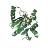
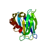
THE ELECTRON DENSITY MAP CONTAINS MANY BLOBS AND CONTINUOUS NON-INTERPRETABLE PATCHES OF DENSITY, ...THE ELECTRON DENSITY MAP CONTAINS MANY BLOBS AND CONTINUOUS NON-INTERPRETABLE PATCHES OF DENSITY, POSSIBLY OWING TO DISORDERED REGIONS INCLUDING SHORT POLYPEPTIDES PRODUCED DURING CRYSTALLISATION UNDER PROTEOLYTIC CONDITIONS. IN PARTICULAR, THE DENSITY IN THE REGION BETWEEN TRP28 AND GLU34 COULD NOT BE INTERPRETED IN TERMS OF ATOMIC MODEL BUT HAS FEATURES SUGGESTING THE PRESENCE OF CHAIN FRAGMENTS CLEAVED AT ARG33 AND OVERLAPPING WITH THEMSELVES AND, POSSIBLY, WITH THE COMPONENTS OF CRYSTALLIZATION SOLUTION. SIMILARLY, A WEAK BUT CONTINUOUS ELECTRON DENSITY BEYOND VAL81 AND LEU141 COULD NOT BE RELIABLY INTERPRETED.
-
Experimental details
-
Experiment
Experiment
Method: X-RAY DIFFRACTION / Number of used crystals: 1
-
Sample preparation
Crystal
Density Matthews: 2.08 Å3/Da / Density % sol: 44 %
Crystal grow
Temperature: 293 K / Method: vapor diffusion / pH: 10.5 Details: Sodium-potassium-phosphate, sodium citrate and CAPS buffer
-
Data collection
Diffraction
Mean temperature: 100 K
Diffraction source
Source: SYNCHROTRON / Site: PETRA III, EMBL c/o DESY / Beamline: P13 (MX1) / Wavelength: 0.97858 Å
Method to determine structure: MOLECULAR REPLACEMENT Starting model: INCOMPLETE MODEL OF SE-MET VARIANT SOLVED BY SAD METHOD USING CRANK2 SOFTWARE Resolution: 2.4→63.03 Å / Cor.coef. Fo:Fc: 0.961 / Cor.coef. Fo:Fc free: 0.948 / SU B: 16.623 / SU ML: 0.179 / Cross valid method: THROUGHOUT / ESU R: 0.345 / ESU R Free: 0.229 / Stereochemistry target values: MAXIMUM LIKELIHOOD / Details: HYDROGENS HAVE BEEN ADDED IN THE RIDING POSITIONS
Rfactor
Num. reflection
% reflection
Selection details
Rfree
0.22364
298
4.8 %
RANDOM
Rwork
0.18623
-
-
-
obs
0.18806
5904
97.21 %
-
Solvent computation
Ion probe radii: 0.8 Å / Shrinkage radii: 0.8 Å / VDW probe radii: 1.2 Å / Solvent model: MASK
 Movie
Movie Controller

 Yorodumi
 Open data
 Basic information
 Components
 Keywords
 Function and homology information
 Ogataea polymorpha (fungus)
 X-RAY DIFFRACTION /
  Authors
 Citation
 Structure visualization
 Downloads & links
 Other downloads

 PDBj
PDBj
 Assembly
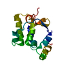

 Mass: 18.015 Da / Num. of mol.: 6 / Source method: isolated from a natural source / Formula: H2O
 Sample preparation
 / Beamline: P13 (MX1) / Wavelength: 0.97858 Å
 Processing