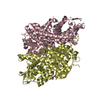
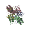
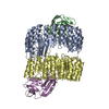
- PDB-5npl: Crystal structure of hexameric CBS-CP12 protein from bloom-formin... -
+
Open data
ID or keywords:
Loading...
-
Basic information
Entry
Database: PDB / ID: 5npl
Title
Crystal structure of hexameric CBS-CP12 protein from bloom-forming cyanobacteria, Yb-derivative at 2.8 A resolution
Components
Similar to tr|Q8YYT1|Q8YYT1
Keywords
PHOTOSYNTHESIS / Cystathionine beta synthase domain / fusion protein / redox-regulation of photosynthesis
Function / homology
Function and homology information
CP12 domain / CP12 domain / CP12 / : / Domain in cystathionine beta-synthase and other proteins. / CBS domain superfamily / CBS domain / CBS domain / CBS domain profile. Similarity search - Domain/homology
Mass: 18.015 Da / Num. of mol.: 51 / Source method: isolated from a natural source / Formula: H2O
Has protein modification
Y
Sequence details
Un-modelled atoms are due to lack of electron density support.
-
Experimental details
-
Experiment
Experiment
Method: X-RAY DIFFRACTION / Number of used crystals: 1
-
Sample preparation
Crystal
Density Matthews: 2.13 Å3/Da / Density % sol: 44 %
Crystal grow
Temperature: 292 K / Method: vapor diffusion / pH: 8 Details: 300 MM NA ACETATE, 20% PEG2000 MME, REMARK 280 100 MM HEPES BUFFER, PH 8.0; before data collection crystals were soaked for 30 min in a solution containing 25% PEG2000MME, 0.2 M sodium ...Details: 300 MM NA ACETATE, 20% PEG2000 MME, REMARK 280 100 MM HEPES BUFFER, PH 8.0; before data collection crystals were soaked for 30 min in a solution containing 25% PEG2000MME, 0.2 M sodium acetate, 0.1 M HEPES, pH 8.0 and 0.1 M Yb-HPDO3A
-
Data collection
Diffraction
Mean temperature: 100 K
Diffraction source
Source: SYNCHROTRON / Site: PETRA III, EMBL c/o DESY / Beamline: P13 (MX1) / Wavelength: 1.385 Å
Detector
Type: DECTRIS PILATUS3 S 6M / Detector: PIXEL / Date: Nov 10, 2015 / Details: mirrors
Radiation
Protocol: SINGLE WAVELENGTH / Monochromatic (M) / Laue (L): M / Scattering type: x-ray
Radiation wavelength
Wavelength: 1.385 Å / Relative weight: 1
Reflection
Resolution: 2.79→66 Å / Num. obs: 13464 / % possible obs: 97.8 % / Redundancy: 6.5 % / Biso Wilson estimate: 51.1 Å2 / Rmerge(I) obs: 0.095 / Net I/σ(I): 14.5
Reflection shell
Resolution: 2.79→2.87 Å / Redundancy: 2.7 % / Rmerge(I) obs: 0.63 / Mean I/σ(I) obs: 1.77 / % possible all: 61.2
-
Processing
Software
Name
Version
Classification
REFMAC
5.8.0155
refinement
XDS
datareduction
XDS
datascaling
CRANK2
phasing
Refinement
Method to determine structure: SAD / Resolution: 2.79→10 Å / Cor.coef. Fo:Fc: 0.942 / Cor.coef. Fo:Fc free: 0.926 / SU B: 25.935 / SU ML: 0.259 / Cross valid method: THROUGHOUT / ESU R Free: 0.359 / Stereochemistry target values: MAXIMUM LIKELIHOOD / Details: HYDROGENS HAVE BEEN ADDED IN THE RIDING POSITIONS
Rfactor
Num. reflection
% reflection
Selection details
Rfree
0.21948
677
4.9 %
RANDOM
Rwork
0.18593
-
-
-
obs
0.18764
13151
95.52 %
-
Solvent computation
Ion probe radii: 0.8 Å / Shrinkage radii: 0.8 Å / VDW probe radii: 1.2 Å / Solvent model: MASK
 Movie
Movie Controller

 Yorodumi
 Open data
 Basic information
 Components
 Keywords
 Function and homology information
 Microcystis aeruginosa PCC 7806 (bacteria)
 X-RAY DIFFRACTION /
  Authors
 Citation
 Structure visualization
 Downloads & links
 Other downloads
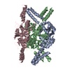


 PDBj
PDBj

 Assembly

 Mass: 173.040 Da / Num. of mol.: 17 / Source method: obtained synthetically / Formula: Yb
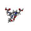
 Mass: 404.459 Da / Num. of mol.: 2 / Source method: obtained synthetically / Formula: C17H32N4O7
 Mass: 18.015 Da / Num. of mol.: 51 / Source method: isolated from a natural source / Formula: H2O
 Sample preparation
 / Beamline: P13 (MX1) / Wavelength: 1.385 Å
 Processing