 Movie
Movie Controller

[English] 日本語
 Yorodumi
- PDB-5nmu: Structure of hexameric CBS-CP12 protein from bloom-forming cyanob... -
+ Open data

- Basic information
| Entry | Database: PDB / ID: 5nmu | ||||||
|---|---|---|---|---|---|---|---|
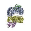
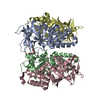
| Title | Structure of hexameric CBS-CP12 protein from bloom-forming cyanobacteria | ||||||
 Components Components | CBS-CP12 | ||||||
 Keywords Keywords | PHOTOSYNTHESIS / Cystathionine beta synthase domain / fusion protein / redox-regulation of photosynthesis | ||||||
| Function / homology |  Function and homology information Function and homology informationCP12 domain / CP12 domain / CP12 / : / Domain in cystathionine beta-synthase and other proteins. / CBS domain superfamily / CBS domain / CBS domain / CBS domain profile. Similarity search - Domain/homology | ||||||
| Biological species |  Microcystis aeruginosa PCC 7806 (bacteria) Microcystis aeruginosa PCC 7806 (bacteria) | ||||||
| Method |  X-RAY DIFFRACTION / SYNCHROTRON / FOURIER SYNTHESIS / Resolution: 2.15 Å X-RAY DIFFRACTION / SYNCHROTRON / FOURIER SYNTHESIS / Resolution: 2.15 Å | ||||||
 Authors Authors | Hackenberg, C. / Hakanpaa, J. / Antonyuk, S.V. / Dittmann, E. / Lamzin, V.S. | ||||||
 Citation Citation | Journal: Proc. Natl. Acad. Sci. U.S.A. / Year: 2018 Title: Structural and functional insights into the unique CBS-CP12 fusion protein family in cyanobacteria. Authors: Hackenberg, C. / Hakanpaa, J. / Cai, F. / Antonyuk, S. / Eigner, C. / Meissner, S. / Laitaoja, M. / Janis, J. / Kerfeld, C.A. / Dittmann, E. / Lamzin, V.S. | ||||||
| History |
|
- Structure visualization
| Structure viewer | Molecule: MolmilJmol/JSmol |
|---|
- Downloads & links
-Download
| PDBx/mmCIF format | 5nmu.cif.gz | 216.2 KB | Display | PDBx/mmCIF format |
|---|---|---|---|---|
| PDB format | pdb5nmu.ent.gz | 174.3 KB | Display | PDB format |
| PDBx/mmJSON format | 5nmu.json.gz | Tree view | PDBx/mmJSON format | |
| Others |  Other downloads Other downloads |
-Validation report
| Arichive directory | https://data.pdbj.org/pub/pdb/validation_reports/nm/5nmuftp://data.pdbj.org/pub/pdb/validation_reports/nm/5nmu | HTTPS FTP |
|---|
-Related structure data
| Related structure data | 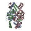 5nplSC  5nvdC S: Starting model for refinement C: citing same article ( |
|---|---|
| Similar structure data |


-Links
 PDBj
PDBj
- Assembly
| Deposited unit | 
| ||||||||
|---|---|---|---|---|---|---|---|---|---|
| 1 | 
| ||||||||
| Unit cell |
|
-Components
| #1: Protein | Mass: 23154.561 Da / Num. of mol.: 3 Source method: isolated from a genetically manipulated source Details: Extra residues are left after TAG cleavage Source: (gene. exp.) Microcystis aeruginosa PCC 7806 (bacteria)Gene: IPF_2164 / Production host: #2: Chemical | ChemComp-CL /   Mass: 35.453 Da / Num. of mol.: 4 / Source method: obtained synthetically / Formula: Cl Mass: 35.453 Da / Num. of mol.: 4 / Source method: obtained synthetically / Formula: Cl#3: Water | ChemComp-HOH / |  Mass: 18.015 Da / Num. of mol.: 191 / Source method: isolated from a natural source / Formula: H2O Mass: 18.015 Da / Num. of mol.: 191 / Source method: isolated from a natural source / Formula: H2OHas protein modification | Y | |
|---|
-Experimental details
-Experiment
| Experiment | Method: X-RAY DIFFRACTION / Number of used crystals: 1 |
|---|
- Sample preparation
| Crystal | Density Matthews: 2.13 Å3/Da / Density % sol: 44 % / Description: hanging drop |
|---|---|
| Crystal grow | Temperature: 292 K / Method: vapor diffusion / pH: 8 Details: 300 mM Na Acetate, 25% PEG2000 MME, 100 mM HEPES buffer, pH 8.0, Vapor diffusion, Temperature 292K |
-Data collection
| Diffraction | Mean temperature: 100 K |
|---|---|
| Diffraction source | Source: SYNCHROTRON / Site: PETRA III, EMBL c/o DESY  / Beamline: P13 (MX1) / Wavelength: 0.976 Å / Beamline: P13 (MX1) / Wavelength: 0.976 Å |
| Detector | Type: DECTRIS PILATUS 6M / Detector: PIXEL / Date: Oct 17, 2015 / Details: KB mirrors |
| Radiation | Monochromator: double crystal monochromator / Protocol: SINGLE WAVELENGTH / Monochromatic (M) / Laue (L): M / Scattering type: x-ray |
| Radiation wavelength | Wavelength: 0.976 Å / Relative weight: 1 |
| Reflection | Resolution: 2.15→68.7 Å / Num. obs: 32084 / % possible obs: 97.6 % / Observed criterion σ(F): 0 / Observed criterion σ(I): 0 / Redundancy: 3.4 % / Biso Wilson estimate: 39.7 Å2 / CC1/2: 0.999 / Rmerge(I) obs: 0.039 / Rpim(I) all: 0.038 / Net I/σ(I): 14.8 |
| Reflection shell | Resolution: 2.15→2.22 Å / Redundancy: 3.5 % / Rmerge(I) obs: 0.384 / Mean I/σ(I) obs: 2.5 / Num. unique obs: 2725 / CC1/2: 0.811 / Rpim(I) all: 0.366 / % possible all: 96.3 |
- Processing
| Software |
| ||||||||||||||||||||||||||||||||||||||||||||||||||||||||||||||||||||||||||||||||||||||||||||||||||||||||||||||||||||||||||||||||||||||||||||||||||||||||||||||||||||||||||||||||||||||
|---|---|---|---|---|---|---|---|---|---|---|---|---|---|---|---|---|---|---|---|---|---|---|---|---|---|---|---|---|---|---|---|---|---|---|---|---|---|---|---|---|---|---|---|---|---|---|---|---|---|---|---|---|---|---|---|---|---|---|---|---|---|---|---|---|---|---|---|---|---|---|---|---|---|---|---|---|---|---|---|---|---|---|---|---|---|---|---|---|---|---|---|---|---|---|---|---|---|---|---|---|---|---|---|---|---|---|---|---|---|---|---|---|---|---|---|---|---|---|---|---|---|---|---|---|---|---|---|---|---|---|---|---|---|---|---|---|---|---|---|---|---|---|---|---|---|---|---|---|---|---|---|---|---|---|---|---|---|---|---|---|---|---|---|---|---|---|---|---|---|---|---|---|---|---|---|---|---|---|---|---|---|---|---|
| Refinement | Method to determine structure: FOURIER SYNTHESIS Starting model: 5NPL Resolution: 2.15→38.58 Å / Cor.coef. Fo:Fc: 0.966 / Cor.coef. Fo:Fc free: 0.94 / SU B: 11.801 / SU ML: 0.15 / Cross valid method: THROUGHOUT / ESU R: 0.218 / ESU R Free: 0.192 / Details: HYDROGENS HAVE BEEN ADDED IN THE RIDING POSITIONS
| ||||||||||||||||||||||||||||||||||||||||||||||||||||||||||||||||||||||||||||||||||||||||||||||||||||||||||||||||||||||||||||||||||||||||||||||||||||||||||||||||||||||||||||||||||||||
| Solvent computation | Ion probe radii: 0.8 Å / Shrinkage radii: 0.8 Å / VDW probe radii: 1.2 Å | ||||||||||||||||||||||||||||||||||||||||||||||||||||||||||||||||||||||||||||||||||||||||||||||||||||||||||||||||||||||||||||||||||||||||||||||||||||||||||||||||||||||||||||||||||||||
| Displacement parameters | Biso mean: 53.779 Å2
| ||||||||||||||||||||||||||||||||||||||||||||||||||||||||||||||||||||||||||||||||||||||||||||||||||||||||||||||||||||||||||||||||||||||||||||||||||||||||||||||||||||||||||||||||||||||
| Refinement step | Cycle: 1 / Resolution: 2.15→38.58 Å
| ||||||||||||||||||||||||||||||||||||||||||||||||||||||||||||||||||||||||||||||||||||||||||||||||||||||||||||||||||||||||||||||||||||||||||||||||||||||||||||||||||||||||||||||||||||||
| Refine LS restraints |
|