 Movie
Movie Controller


+ Open data

- Basic information
| Entry | Database: PDB / ID: 1a4y | ||||||
|---|---|---|---|---|---|---|---|
| Title | RIBONUCLEASE INHIBITOR-ANGIOGENIN COMPLEX | ||||||
 Components Components |
| ||||||
 Keywords Keywords | COMPLEX (INHIBITOR/NUCLEASE) / COMPLEX (INHIBITOR-NUCLEASE) / COMPLEX (RI-ANG) / HYDROLASE MOLECULAR RECOGNITION / EPITOPE MAPPING / LEUCINE-RICH REPEATS / COMPLEX (INHIBITOR-NUCLEASE) complex | ||||||
| Function / homology |  Function and homology information Function and homology informationribonuclease inhibitor activity / tRNA stabilization / angiogenin-PRI complex / tRNA-specific ribonuclease activity / negative regulation of translation in response to stress / tRNA-derived small RNA (tsRNA or tRNA-related fragment, tRF) biogenesis / tRNA decay / regulation of Arp2/3 complex-mediated actin nucleation / signaling / cell communication ...ribonuclease inhibitor activity / tRNA stabilization / angiogenin-PRI complex / tRNA-specific ribonuclease activity / negative regulation of translation in response to stress / tRNA-derived small RNA (tsRNA or tRNA-related fragment, tRF) biogenesis / tRNA decay / regulation of Arp2/3 complex-mediated actin nucleation / signaling / cell communication / oocyte maturation / Hydrolases; Acting on ester bonds; Endoribonucleases producing 3'-phosphomonoesters / Adherens junctions interactions / hematopoietic stem cell proliferation / mRNA catabolic process / homeostatic process / rRNA transcription / basement membrane / positive regulation of phosphorylation / regulation of angiogenesis / RNA nuclease activity / endocytic vesicle / ovarian follicle development / actin filament polymerization / peptide binding / positive regulation of endothelial cell proliferation / RNA endonuclease activity / response to hormone / placenta development / stress granule assembly / positive regulation of protein secretion / negative regulation of smooth muscle cell proliferation / cytoplasmic stress granule / antimicrobial humoral immune response mediated by antimicrobial peptide / cell migration / heparin binding / lamellipodium / actin cytoskeleton / chromosome / antibacterial humoral response / growth cone / ribosome binding / endonuclease activity / actin binding / angiogenesis / response to hypoxia / defense response to Gram-positive bacterium / rRNA binding / receptor ligand activity / copper ion binding / signaling receptor binding / innate immune response / hydrolase activity / neuronal cell body / negative regulation of apoptotic process / nucleolus / signal transduction / protein homodimerization activity / : / DNA binding / extracellular exosome / extracellular region / nucleoplasm / nucleus / plasma membrane / cytosol / cytoplasm Similarity search - Function | ||||||
| Biological species |  Homo sapiens (human) Homo sapiens (human) | ||||||
| Method |  X-RAY DIFFRACTION / SYNCHROTRON / MOLECULAR REPLACEMENT / Resolution: 2 Å X-RAY DIFFRACTION / SYNCHROTRON / MOLECULAR REPLACEMENT / Resolution: 2 Å | ||||||
 Authors Authors | Papageorgiou, A.C. / Acharya, K.R. | ||||||
 Citation Citation | Journal: EMBO J. / Year: 1997 Title: Molecular recognition of human angiogenin by placental ribonuclease inhibitor--an X-ray crystallographic study at 2.0 A resolution. Authors: Papageorgiou, A.C. / Shapiro, R. / Acharya, K.R. | ||||||
| History |
|
- Structure visualization
| Structure viewer | Molecule: MolmilJmol/JSmol |
|---|
- Downloads & links
-Download
| PDBx/mmCIF format | 1a4y.cif.gz | 226.4 KB | Display | PDBx/mmCIF format |
|---|---|---|---|---|
| PDB format | pdb1a4y.ent.gz | 181.8 KB | Display | PDB format |
| PDBx/mmJSON format | 1a4y.json.gz | Tree view | PDBx/mmJSON format | |
| Others |  Other downloads Other downloads |
-Validation report
| Arichive directory | https://data.pdbj.org/pub/pdb/validation_reports/a4/1a4yftp://data.pdbj.org/pub/pdb/validation_reports/a4/1a4y | HTTPS FTP |
|---|
-Related structure data
| Related structure data |  1bnh S: Starting model for refinement |
|---|---|
| Similar structure data |


-Links
 PDBj
PDBj


- Assembly
| Deposited unit | 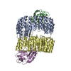
| ||||||||
|---|---|---|---|---|---|---|---|---|---|
| 1 |
| ||||||||
| Unit cell |
| ||||||||
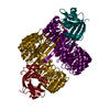
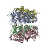
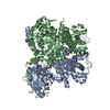
| Noncrystallographic symmetry (NCS) | NCS oper: (Code: given Matrix: (-0.63908, 0.58117, 0.50381), Vector: Details | THERE ARE TWO COMPLEXES IN THE ASYMMETRIC UNIT (CHAINS A, B, C AND D, E, F). CHAINS A AND D CORRESPOND TO THE INHIBITOR, CHAINS B AND E CORRESPOND TO ANGIOGENIN, AND CHAINS C AND F CORRESPOND TO THE WATER MOLECULES ASSOCIATED WITH THE RESPECTIVE COMPLEX. | |
-Components
| #1: Protein | Mass: 49887.156 Da / Num. of mol.: 2 Source method: isolated from a genetically manipulated source Source: (gene. exp.) Homo sapiens (human) / Organ: PLACENTA / Plasmid: PANG2 / Species (production host): Escherichia coliProduction host:  Strain (production host): W3110 / References: UniProt: P13489 #2: Protein | Mass: 14169.036 Da / Num. of mol.: 2 Source method: isolated from a genetically manipulated source Source: (gene. exp.) Homo sapiens (human) / Organ: PLACENTA / Plasmid: PANG2 / Species (production host): Escherichia coliProduction host: Strain (production host): W3110 References: UniProt: P03950, Hydrolases; Acting on ester bonds; Endoribonucleases producing 3'-phosphomonoesters #3: Water | ChemComp-HOH / |  Mass: 18.015 Da / Num. of mol.: 133 / Source method: isolated from a natural source / Formula: H2O Mass: 18.015 Da / Num. of mol.: 133 / Source method: isolated from a natural source / Formula: H2OHas protein modification | Y | |
|---|
-Experimental details
-Experiment
| Experiment | Method: X-RAY DIFFRACTION |
|---|
- Sample preparation
| Crystal | Density Matthews: 2.5 Å3/Da / Density % sol: 49.82 % Description: THE POSITION OF ANGIOGENIN WAS LOCATED BY EXAMINATION OF THE ELECTRON DENSITY MAP | ||||||||||||||||||||||||||||||||||||||||||||||||||||||
|---|---|---|---|---|---|---|---|---|---|---|---|---|---|---|---|---|---|---|---|---|---|---|---|---|---|---|---|---|---|---|---|---|---|---|---|---|---|---|---|---|---|---|---|---|---|---|---|---|---|---|---|---|---|---|---|
| Crystal grow | pH: 4.2 Details: THE COMPLEX WAS CRYSTALLIZED FROM 10% PEG4000, 20MM SODIUM CITRATE (PH 4.2), 0.1 AMMONIUM SULPHATE AND 25 MM DTT. | ||||||||||||||||||||||||||||||||||||||||||||||||||||||
| Crystal | *PLUS | ||||||||||||||||||||||||||||||||||||||||||||||||||||||
| Crystal grow | *PLUS Temperature: 16 ℃ / Method: vapor diffusion, hanging drop / pH: 7.2 | ||||||||||||||||||||||||||||||||||||||||||||||||||||||
| Components of the solutions | *PLUS
|
-Data collection
| Diffraction | Mean temperature: 289 K | |||||||||
|---|---|---|---|---|---|---|---|---|---|---|
| Diffraction source | Source: SYNCHROTRON / Site: SRS  / Beamline: PX9.6 / Wavelength: 0.87 / Wavelength: 0.87, 1.5418 / Beamline: PX9.6 / Wavelength: 0.87 / Wavelength: 0.87, 1.5418 | |||||||||
| Detector | Type: MARRESEARCH / Detector: IMAGE PLATE / Date: Jun 1, 1996 | |||||||||
| Radiation | Monochromatic (M) / Laue (L): M / Scattering type: x-ray | |||||||||
| Radiation wavelength |
| |||||||||
| Reflection | Resolution: 2→40 Å / Num. obs: 72355 / % possible obs: 86.9 % / Observed criterion σ(I): 0 / Redundancy: 2.2 % / Biso Wilson estimate: 40.4 Å2 / Rmerge(I) obs: 0.094 / Net I/σ(I): 4.5 | |||||||||
| Reflection shell | Resolution: 2→2.07 Å / Rmerge(I) obs: 0.436 / Mean I/σ(I) obs: 1.6 / % possible all: 65.4 | |||||||||
| Reflection | *PLUS Num. measured all: 158593 | |||||||||
| Reflection shell | *PLUS Highest resolution: 2 Å / % possible obs: 65.4 % |
- Processing
| Software |
| ||||||||||||||||||||||||||||||||||||||||||||||||||||||||||||||||||||||||||||||||
|---|---|---|---|---|---|---|---|---|---|---|---|---|---|---|---|---|---|---|---|---|---|---|---|---|---|---|---|---|---|---|---|---|---|---|---|---|---|---|---|---|---|---|---|---|---|---|---|---|---|---|---|---|---|---|---|---|---|---|---|---|---|---|---|---|---|---|---|---|---|---|---|---|---|---|---|---|---|---|---|---|---|
| Refinement | Method to determine structure: MOLECULAR REPLACEMENT Starting model: 1BNH (PIG RIBONUCLEASE INHIBITOR) 1bnh Resolution: 2→20 Å / Data cutoff high absF: 1000000 / Data cutoff low absF: 0.001 / Isotropic thermal model: RESTRAINED / Cross valid method: THROUGHOUT / σ(F): 0
| ||||||||||||||||||||||||||||||||||||||||||||||||||||||||||||||||||||||||||||||||
| Displacement parameters | Biso mean: 42 Å2 | ||||||||||||||||||||||||||||||||||||||||||||||||||||||||||||||||||||||||||||||||
| Refine analyze | Luzzati sigma a free: 0.36 Å | ||||||||||||||||||||||||||||||||||||||||||||||||||||||||||||||||||||||||||||||||
| Refinement step | Cycle: LAST / Resolution: 2→20 Å
| ||||||||||||||||||||||||||||||||||||||||||||||||||||||||||||||||||||||||||||||||
| Refine LS restraints |
| ||||||||||||||||||||||||||||||||||||||||||||||||||||||||||||||||||||||||||||||||
| Xplor file |
| ||||||||||||||||||||||||||||||||||||||||||||||||||||||||||||||||||||||||||||||||
| Software | *PLUS Name: X-PLOR / Version: 3.8 / Classification: refinement | ||||||||||||||||||||||||||||||||||||||||||||||||||||||||||||||||||||||||||||||||
| Refinement | *PLUS | ||||||||||||||||||||||||||||||||||||||||||||||||||||||||||||||||||||||||||||||||
| Solvent computation | *PLUS | ||||||||||||||||||||||||||||||||||||||||||||||||||||||||||||||||||||||||||||||||
| Displacement parameters | *PLUS Biso mean: 42 Å2 |
