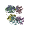
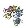
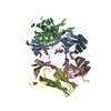
Journal: Proc Natl Acad Sci U S A / Year: 2015 Title: pH-Dependent recognition of apoptotic and necrotic cells by the human dendritic cell receptor DEC205. Authors: Longxing Cao / Xiangyi Shi / Haishuang Chang / Qinfen Zhang / Yongning He / Abstract: Dendritic cells play important roles in regulating innate and adaptive immune responses. DEC205 (CD205) is one of the major endocytotic receptors on dendritic cells and has been widely used for ...Dendritic cells play important roles in regulating innate and adaptive immune responses. DEC205 (CD205) is one of the major endocytotic receptors on dendritic cells and has been widely used for vaccine generation against viruses and tumors. However, little is known about its structure and functional mechanism. Here we determine the structure of the human DEC205 ectodomain by cryoelectron microscopy. The structure shows that the 12 extracellular domains form a compact double ring-shaped conformation at acidic pH and become extended at basic pH. Biochemical data indicate that the pH-dependent conformational change of DEC205 is correlated with ligand binding and release. DEC205 only binds to apoptotic and necrotic cells at acidic pH, whereas live cells cannot be recognized by DEC205 at either acidic or basic conditions. These results suggest that DEC205 is an immune receptor that recognizes apoptotic and necrotic cells specifically through a pH-dependent mechanism.
History
Deposition
May 5, 2015
-
Header (metadata) release
May 27, 2015
-
Map release
May 27, 2015
-
Update
May 25, 2016
-
Current status
May 25, 2016
Processing site: PDBj / Status: Released
-
Structure visualization
Movie
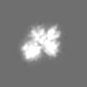
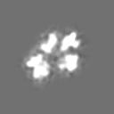
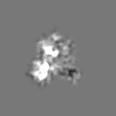
Surface view with section colored by density value
Organism: Homo sapiens (human) / synonym: Human / Location in cell: Plasma membrane
Molecular weight
Theoretical: 191.2 MDa
Recombinant expression
Organism: Homo sapiens (human) / Recombinant strain: Human / Recombinant cell: HEK293 / Recombinant plasmid: pTT5
Sequence
UniProtKB: Lymphocyte antigen 75
-
Experimental details
-
Structure determination
Method
negative staining, cryo EM
Processing
single particle reconstruction
Aggregation state
particle
-
Sample preparation
Concentration
3.0 mg/mL
Buffer
pH: 6 / Details: 150mM NaCl, 50mM Bis-Tris
Staining
Type: NEGATIVE / Details: Cryo
Grid
Details: Quantifoil Holey Carbon Film
Vitrification
Cryogen name: ETHANE / Chamber humidity: 100 % / Chamber temperature: 77 K / Instrument: FEI VITROBOT MARK II / Method: Blot for 6 seconds before plunging
-
Electron microscopy
Microscope
JEOL 2100F
Specialist optics
Energy filter - Name: in column filter
Date
Apr 1, 2012
Image recording
Category: CCD / Film or detector model: GATAN ULTRASCAN 4000 (4k x 4k) / Number real images: 153 / Average electron dose: 20 e/Å2
Electron beam
Acceleration voltage: 200 kV / Electron source: FIELD EMISSION GUN
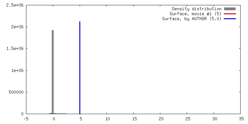
Algorithm: OTHER / Resolution.type: BY AUTHOR / Resolution: 14.6 Å / Resolution method: OTHER / Software - Name: EMAN, EMAN2 / Number images used: 15723
Final two d classification
Number classes: 20
+
About Yorodumi
-
News
-
Feb 9, 2022. New format data for meta-information of EMDB entries
New format data for meta-information of EMDB entries
Version 3 of the EMDB header file is now the official format.
The previous official version 1.9 will be removed from the archive.
In the structure databanks used in Yorodumi, some data are registered as the other names, "COVID-19 virus" and "2019-nCoV". Here are the details of the virus and the list of structure data.
Jan 31, 2019. EMDB accession codes are about to change! (news from PDBe EMDB page)
EMDB accession codes are about to change! (news from PDBe EMDB page)
The allocation of 4 digits for EMDB accession codes will soon come to an end. Whilst these codes will remain in use, new EMDB accession codes will include an additional digit and will expand incrementally as the available range of codes is exhausted. The current 4-digit format prefixed with “EMD-” (i.e. EMD-XXXX) will advance to a 5-digit format (i.e. EMD-XXXXX), and so on. It is currently estimated that the 4-digit codes will be depleted around Spring 2019, at which point the 5-digit format will come into force.
The EM Navigator/Yorodumi systems omit the EMD- prefix.
Related info.:Q: What is EMD? / ID/Accession-code notation in Yorodumi/EM Navigator
Yorodumi is a browser for structure data from EMDB, PDB, SASBDB, etc.
This page is also the successor to EM Navigator detail page, and also detail information page/front-end page for Omokage search.
The word "yorodu" (or yorozu) is an old Japanese word meaning "ten thousand". "mi" (miru) is to see.
Related info.:EMDB / PDB / SASBDB / Comparison of 3 databanks / Yorodumi Search / Aug 31, 2016. New EM Navigator & Yorodumi / Yorodumi Papers / Jmol/JSmol / Function and homology information / Changes in new EM Navigator and Yorodumi
 Movie
Movie Controller


 Open data
 Basic information
 Map data
 Sample
 Keywords
 Function and homology information
 Homo sapiens (human)
 Authors
 Citation
 Structure visualization
 Downloads & links
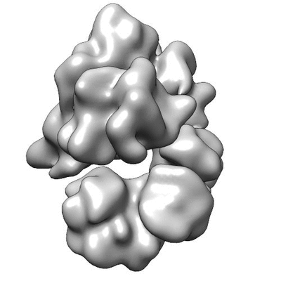 400_6333.gif
 80_6333.gif
 http://ftp.pdbj.org/pub/emdb/structures/EMD-6333


 Z (Sec.)
 Y (Row.)
 X (Col.)


 Sample components
 Processing
 Electron microscopy
 FIELD EMISSION GUN